Introduction
Ototoxicity is the pharmacological adverse reaction which results from exposure to drugs or chemicals that damage the inner ear or the vestibulocochlear nerve [1]. Toxic drug-induced hearing loss is increasing worldwide, and many countries are actively conducting research on this toxic hearing loss [2,3]. Many patients with anticancer therapy or antibiotics for long-term administration have a high incidence of sensorineural hearing loss (SNHL) [4].
Drug-related ototoxic hearing loss has been demonstrated as a bilateral SNHL [5]. The patients with ototoxic hearing loss do not realize when their hearing loss are aggravation. Severity of incidence rate of ototoxic hearing loss shows dose-dependent and cumulative. It can be influenced by various factors; noise, sex, age, and co-morbid diseases [1]. To solve the degenerative change of hair cell, research on hair cell regeneration for bone marrow derived mesenchymal stem cells (BM-MSCs) has been conducted.
BM-MSCs are interesting clinical research source. Stem cells can self-renew with a high growth rate, possess multipotent differentiation properties. In recently, it is also easily accessible and regenerable [6,7]. Adult stem cells also have the potential to deliver gene and therapeutic molecules to other parts of the inner ear [8]. Stem cells can be categorized as fetal stem cells, embryonic stem cells, adult stem cells, induced pluripotent stem cells and perinatal stem cells [9]. Adult stem cells play a key role in tissue regeneration and have multipotent cells present in most adult tissues [4]. Besides, neuronal cells such as Schwann cells (supporting cells) can differentiate from the endogenous stem cells in the inner ear and from MSCs [10].
BM-MSCs have been shown to be effective in various diseases, however, their efficacy of MSCs transplantation in ototoxic hearing loss was not evaluated. In this study, we investigated about the efficacy of clinical grade, pre-made, BM-MSCs in our institute (Catholic MASTER cell) on functional hearing improvement after stem cell transplantation.
Materials and Methods
Catholic MASTER cell
BM-MSCs were purchased from the Catholic Institute of Cell Therapy (CIC, Seoul, Korea). It is human BM-MSC ready to use made in good manufacturing practice in CIC call Catholic MASTER cell. BM-MSCs were cultured in DulbeccoÔÇÖs modified eagle medium (Wisent Bioproducts, St. Bruno, QC, Canada) supplemented with 20% fetal bovine serum (Wisent Bioproducts), penicillin, and streptomycin (Gibco, Carlsbad, CA, USA), and maintained at 37┬░C with 5% CO2.
Formation of deaf animal model
All 6 weeks old male C57/BL6 mice (Orient Bio, Seoul, Korea) were used for the experiment. We divided the animals into two groups (n=5 per group); saline injected control group (left ear is normal hearing, control group) and stem cell injected group (right ear is deafened with ototoxic drugs, MSC-group). We anesthetized the mice with a mixture of xylazine (40 mg/mL; Bayer, Seoul, Korea) and zoletil (60 mg/mL; Virbac, Seoul, Korea) by intramuscular injection. The mouse dissection was performed under the Zeiss Universal S2 surgical microscope (Carl Zeiss, Goettingen, Germany).
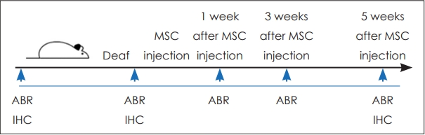
As ototoxic drugs, a mixture of 10% neomycin (SigmaAldrich, St. Louis, MO, USA) and 5 M ouabain octahydrate (Sigma-Aldrich) was injected into the right middle ear with 1 cc syringe to produce the deaf ear. We injected saline into the control group. We also injected stem cells into the MSCgroup via the tail vein using a Hamilton syringe. We administered 1├Ś106 cells in 100 ╬╝L of the BM-MSCs to the experimental group and the same amount of saline was administered to the control group (Fig. 1).
Hearing threshold test
The hearing threshold was evaluated with auditory brainstem response (ABR). The ABR was recorded using sub-dermal needle electrodes at the vertex, below the pinna of the left ear (reference) and below the contralateral ear (ground). The sound stimuli included clicks (100-ms duration; 8 Hz), and ABR measurements were obtained with an Intelligent Hearing System (IHS); the SmartEP System, running the IHS highfrequency software (ver. 2.33; Miami, FL, USA) and using IHS high-frequency transducers. The ABR threshold was measured by detecting the presence of wave V. When the recording intensity showed at least 2 sequences, the response reproducibility was confirmed. ABR was performed at 1, 3, and 5 weeks after the injection of BM-MSCs and saline.
Immunohistochemistry of organ of corti
Mouse was put under anesthesia and the temporal bone was isolated. The cochlea was isolated after removal of temporal bone. The cochlea was perfused with 4% paraformaldehyde and retained in fixative overnight at 4┬░C for the surface preparation. After washing with phosphate buffered saline, the cochlea was decalcified for 3 days. The ReissnerÔÇÖs membrane and tectorial membrane was exposed. The organ of corti (OC) was stained with myosin VIIA (1:200) overnight. The hair cells in the OC was visualized under a microscope equipped with epifluorescence. Fluorescent image was materalized using a Zeiss LSM510 META confocal microscope (Carl Zeiss). The captured images processed using Zen 2009 Light Edition (Carl Zeiss).
Ethics statement
Institutional Animal Care and Use Committee of the Department of Laboratory Animals at the School of Medicine, Catholic University of Korea approved this study and all mice were treated in accordance with the guidelines for the Care and Use of Laboratory Animals of Catholic University College of Medicine (approval No. 2017-0296-02).
Statistical analysis
All statistical analyses were performed using SPSS software for PC, version 21 (IBM Corp., Armonk, NY, USA). The results are presented as mean┬▒SD. Continuous variables were compared using the StudentÔÇÖs t-test for evaluating differences between unpaired groups. For all statistical tests, a p-value of <0.05 was considered as statistically significant.
Results
Hearing recovery after mesenchymal stem cell injection
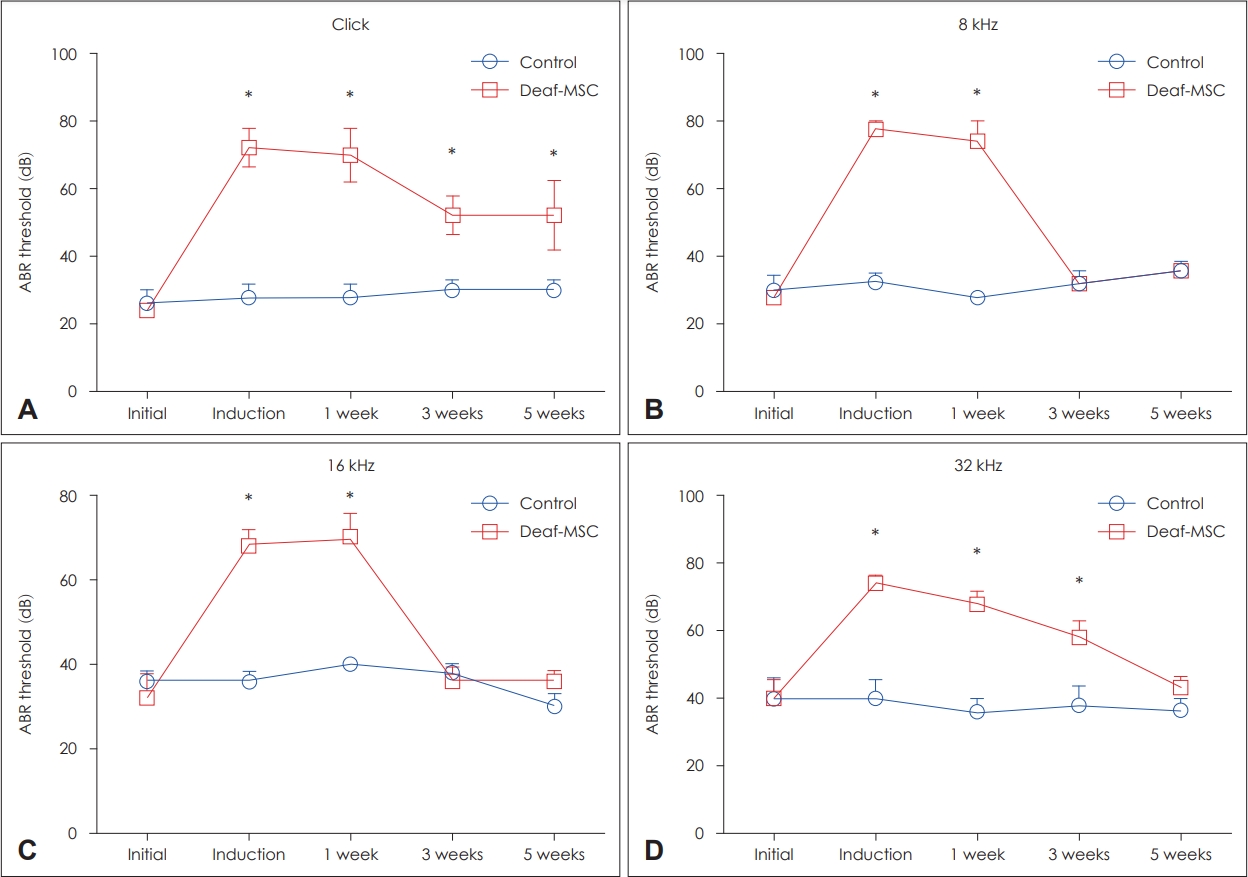
We evaluated the hearing recovery in deaf ear after mesenchymal stem cell injection. Hearing thresholds were evaluated at initial, induction, injection after 1, 3, and 5 weeks with ABR test. The measured hearing threshold of MSC-group after click stimulation was: initial, 24┬▒2.4 dB; induction, 72┬▒5.8 dB; 1 week, 70┬▒7.7 dB; 3 weeks, 52┬▒5.8 dB; and 5 weeks, 52┬▒10.2 dB. The ABR threshold in the MSC-group was significantly higher compared with control ear of the initial, 1, 3, and 5 weeks (p<0.05) (Fig. 2A). The measured hearing threshold of MSC-group after 8 kHz stimulation was: initial, 28┬▒2.0 dB; induction, 78┬▒2.5 dB; 1 week, 74┬▒6.0 dB; 3 weeks, 32┬▒2.0 dB; and 5 weeks, 36┬▒2.4 dB. The ABR threshold in the MSC-group was significantly higher compared with control ear of the initial and 1 week (p<0.05) (Fig. 2B). The measured hearing threshold of MSC-group after 16 kHz stimulation was: initial, 32┬▒5.8 dB; induction, 68┬▒3.7 dB; 1 week, 70┬▒5.4 dB; 3 weeks, 36┬▒4.0 dB; and 5 weeks, 36┬▒2.4 dB. The ABR threshold in the MSC-group was significantly higher compared with control ear of the initial and 1 week (p<0.05) (Fig. 2C). The measured hearing threshold of MSC-group after 32 kHz stimulation was: initial, 40┬▒5.4 dB; induction, 74┬▒5.4 dB; 1 week, 68┬▒3.7 dB; 3 weeks, 58┬▒4.9 dB; and 5 weeks, 43┬▒3.3 dB. The ABR threshold in the MSC-group was significantly higher compared with control ear of the initial, 1 and 3 weeks (p<0.05) (Fig. 2D).
Morphological regeneration following stem cell transplantation in deaf ear
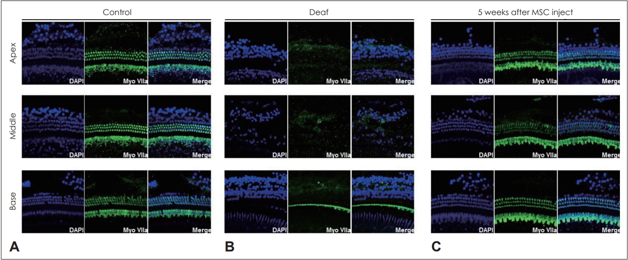
In order to confirm the morphologic restoration of OC after stem cell transplantation, regeneration of OC in MSC-group was compared with control group in 5 weeks after stem cell transplantation. The hair cells of control group showed normal feature after stained with myosin VIIA (Fig. 3A). In the MSC-group, multiple hair cells of apical to the basal turn of the cochlea were destructed (Fig. 3B). Five weeks after transplantation of MSCs, hair cells regeneration was confirmed from the basal turn to the apex of the cochlea in MSC-group (Fig. 3C).
Discussion
This study identifies the effect of BM-MSCs in ototoxic hearing loss with direct functional and molecular evidence. The most salient observations of this study are: 1) Ototoxic hearing loss was functionally recovered by MSCs injection 2) OC was actually restored by MSCs injection in the ototoxic hearing loss model. Therefore, MSCs is highly considered to be involved in hair cell regeneration.
There are about 15000 hair cells in the cochlea which are important for sound sensation [11]. The OC cannot be restored after degeneration. The degeneration of hair cells leads to morphological degeneration of near hair cells [12]. Hair cells are vulnerable and easily damaged by excessive stimulation such as ototoxic drugs and by the natural effects of aging [13]. The cumulative damage causes progressive and permanent hearing loss to the OC.
Previously, we confirmed the effect of transplantation of human umbilical cord blood mesenchymal stem cells (UCB-MSCs) on hearing restoration in guinea pig model with sensorineural hearing loss [6]. The UCB-MSCs group showed a significant hearing restoration after transplantation. The spiral ganglion and hair cells were increased in UCB-MSCs group. Previously, we also evaluated regenerative effect of hearing loss by transplanting mesenchymal stromal cells derived from human placenta in deaf animal model. In the study, BM-MSCs transplantation resulted in auditory improvement and cell regeneration [14]. The ABR results through at 1, 3, and 5 weeks showed the hearing improvement in the MSC-group. In particular, hearing threshold was restored to normal levels at specific frequency at 5 weeks after transplantation (8 khz and 16 khz).
BM-MSCs have been presented as a possible alternative therapy in various pathologies of the neural system. They can replace the injured neural tissue, therefore capable to be an alternative treatment of neural injuries. There were a lot studies that have proven the functional improvements of nervous system degeneration after treatment of BM-MSCs in animal model [9].
In some studies, the possibility of treatment of degenerative hearing loss with stem cell transplantation has been evaluated. Hakuba, et al. [15] reported that the neural stem cell transplantation was useful for preventing injury to hair cells which developed after cochlea transient ischemia. Matsuoka, et al. [16] reported that the survival of transplanted MSCs into the modiolus of the cochlea may result in regeneration of damaged spiral ganglion neurons. These findings have important clinical implications as a means of delivering MSCs in the cochlea for stem-cell replacement therapy. Jeon, et al. [17] suggested that BM-MSCs can differentiate into hair cells and various neurons. Cho, et al. [18] evaluated to confirm the efficacy of transplanted neural differentiated human mesenchymal stem cells (hMSCs). They used the guinea pig model and they showed auditory neuropathy. The study suggests that the stem cells were grafted into the scala tympani, the degenerated SGNs were replaced.
Even in response to usual stimulation occurs in daily life, the OC which is primary site for hearing sensation, can be damaged and cause hearing disorders. Our experiments demonstrated that MSC injection has several possibilities for treating ototoxic hearing loss by restoring the hair cells of the inner ear. We believe that this study provide a basis for treating degenerative hearing disease and reinforce the conviction of Catholic MASTER cell have positive potential for differentiating into inner ear organs in vitro.
This is the first study to demonstrate that Catholic MASTER cell treatment induces hearing recovery and hair cell regeneration in the OC of mouse. The results of this study can justify the role of stem cell transplantation mediated inner ear regeneration.