ņä£ ļĪĀ
Ļ░æņāüņäĀ ņŚ¼ĒżņĢöņØĆ ņĀäņ▓┤ Ļ░æņāüņäĀņĢö ņżæ ļæÉ ļ▓łņ¦ĖļĪ£ ĒØöĒĢ£ ņ£ĀĒśĢņØ┤ļŗż. Ļ░æņāüņäĀņĢö ņżæ ņĢĮ 10%ļź╝ ņ░©ņ¦ĆĒĢśļ®░ ņŻ╝ļĪ£ 50ļīĆņŚÉ ĒØöĒĢśĻ│Ā ņŚ¼ņä▒ņŚÉņä£ ļé©ņä▒ļ│┤ļŗż 3ļ░░ ļŹö ļ¦ÄņØ┤ ļ░£Ļ▓¼ļÉ£ļŗż. Ļ░æņāüņäĀ ņŚ¼ĒżņĢöņØĆ ĒŖ╣Ē׳ ņÜöņśżļō£ļź╝ ņäŁņĘ©ĒĢśĻĖ░ ņ¢┤ļĀżņÜ┤ ņ¦ĆņŚŁņŚÉņä£ ļ¦ÄņØ┤ ļ░£ņāØĒĢśļŖö Ļ▓āņ£╝ļĪ£ ņĢīļĀżņĀĖ ņ׳ļŗż. Ļ░æņāüņäĀ ņŚ¼ĒżņĢöņØĆ ļīĆļČĆļČä ļ¼┤ņ”Øņāüņ£╝ļĪ£ ņÜ░ņŚ░Ē׳ ļ░£Ļ▓¼ļÉ£ Ļ░æņāüņäĀņØś ņóģĻ┤┤ļĪ£ ļ░£ĒśäļÉ£ļŗż. Ļ░æņāüņäĀ ņŚ¼ĒżņĢöņØś ļ”╝ĒöäņĀł ņĀäņØ┤ļŖö 10% ļ»Ėļ¦īņ£╝ļĪ£ ņ£ĀļæÉņĢöņŚÉ ļ╣äĒĢ┤ ņāüļīĆņĀüņ£╝ļĪ£ ļé«ļŗż. ņŚ¼ĒżņĢöņØĆ ņŻ╝ļĪ£ ĒśłĒ¢ēņä▒ ņĀäņØ┤ļź╝ ĒĢśļ®░ ņøÉĻ▓® ņĀäņØ┤ņ£©ņØĆ ņĢĮ 20%ļĪ£ Ļ│© ņĀäņØ┤Ļ░Ć Ļ░Ćņן ĒØöĒĢśļ®░ ļŗżņØīņ£╝ļĪ£ ņ×ÉņŻ╝ ņĀäņØ┤ļÉśļŖö ņןĻĖ░ļŖö Ļ░ä, ĒÅÉ, ļćīņØ┤ļŗż. ņŗĀņןņØś ņóģņ¢æņŚÉņä£ Ļ░æņāüņäĀ ņŚ¼ĒżņĢöņØ┤ ļ©╝ņĀĆ ņ¦äļŗ©ļÉśļŖö Ļ▓ĮņÜ░ļŖö ļ¦żņÜ░ ļō£ļ¼╝ļŗż. ļ│Ė ņ”ØļĪĆļŖö ņŗĀņןņØś ņóģĻ┤┤ļź╝ ņĀ£Ļ▒░ĒĢ£ Ēøä ļ│æļ”¼Ļ▓Ćņé¼ņŚÉņä£ ņŗĀņןņ£╝ļĪ£ ņĀäņØ┤ļÉ£ Ļ░æņāüņäĀ ņŚ¼ĒżņĢöņØ┤ ņ¦äļŗ©ļÉśņ¢┤ Ļ░æņāüņäĀ ņĀäņĀłņĀ£ņłĀņØä ĒåĄĒĢ┤ ņøÉļ░£ļČĆņ£äļź╝ ĒÖĢņØĖĒĢ£ ņ”ØļĪĆļź╝ Ļ▓ĮĒŚśĒĢśņśĆĻĖ░ņŚÉ ļ¼ĖĒŚī Ļ│Āņ░░Ļ│╝ ĒĢ©Ļ╗ś ļ│┤Ļ│ĀĒĢśļŖö ļ░öņØ┤ļŗż.
ņ”Ø ļĪĆ
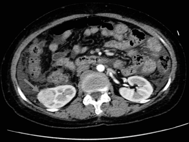
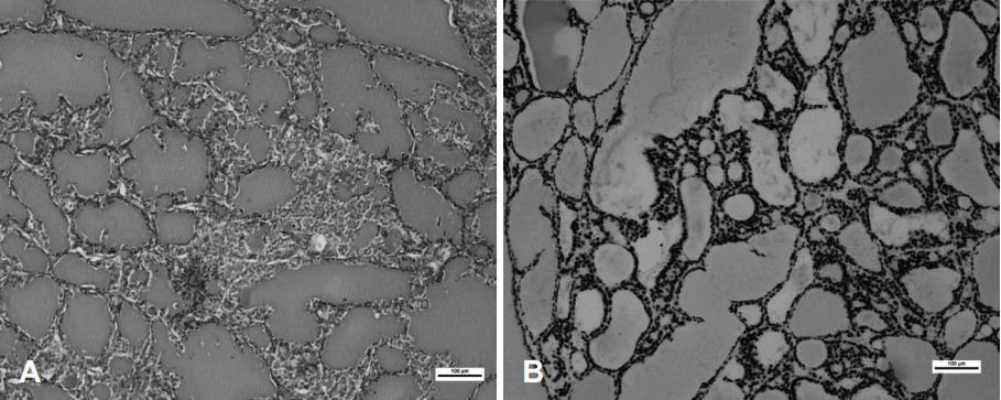
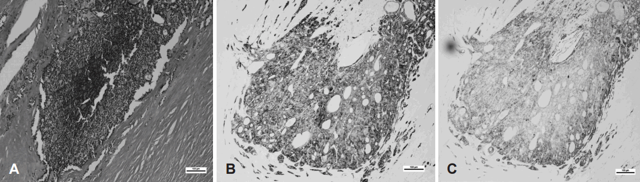
57ņäĖ ņŚ¼ņ×É ĒÖśņ×ÉĻ░Ć ļé┤ņøÉ ņØ╝ņŻ╝ņØ╝ ņĀäļČĆĒä░ ļ░£ņāØĒĢ£ ļ│ĄļČĆļČłĒÄĖĻ░ÉĻ│╝ ļČĆņóģņØä ņŻ╝ņåīļĪ£ ņØæĻĖēņŗżņØä ļé┤ņøÉĒĢśņśĆļŗż. Ļ│╝Ļ▒░ļĀźņāü 20ļģä ņĀä BĒśĢĻ░äņŚ╝ņØä ņ¦äļŗ©ļ░øņĢśņ£╝ļéś ņ╣śļŻīļŖö ļ░øņ¦Ć ņĢŖĻ│Ā ņ׳ņŚłĻ│Ā ņĄ£ĻĘ╝ņŚÉ ĒĢ£ņĢĮņØä ļ│ĄņÜ®ĒĢ£ņĀü ņ׳ņ¢┤ Ļ░äĻ▓ĮĒÖö ņØśņ”Øņ£╝ļĪ£ ļ│ĖņøÉ ņåīĒÖöĻĖ░ ļé┤Ļ│╝ Ēøä ņ╗┤Ēō©Ēä░ļŗ©ņĖĄņ┤¼ņśüņØä ņŗ£Ē¢ēĒĢśņśĆĻ│Ā, ņÜ░ņĖĪ ņŗĀņןņØś ĒĢśļČĆņŚÉņä£ 3.5 cm Ļ░Ćļ¤ēņØś ņóģņ¢æņØ┤ ļ░£Ļ▓¼ļÉśņŚłļŗż(Fig. 1). ņØ┤Ēøä ĒÖśņ×ÉļŖö ņŗĀņן ņäĖĒż ņĢöņóģ(renal cell carcinoma) ņØśņŗ¼ ĒĢśņŚÉ ņÜ░ņĖĪ ĻĘ╝ņ╣śņĀü ņŗĀņןņĀłņĀ£ņłĀņØä ņŗ£Ē¢ēĒĢśņśĆĻ│Ā 3.2├Ś3 cmņØĖ Ļ│ĀĒśĢņä▒ņØś ņŚ░ļ»ĖņāēņØä ļØĀĻ│Ā ņ׳ļŖö ņóģĻ┤┤Ļ░Ć ļ░£Ļ▓¼ļÉśņŚłļŗż. ņĪ░ņ¦üļ│æļ”¼ĒĢÖņĀüņ£╝ļĪ£ hematoxylin & eosin ņŚ╝ņāē ņĪ░ņ¦üĒæ£ļ│ĖņŚÉņä£ ņĮ£ļĪ£ņØ┤ļō£ļź╝ ĒżĒĢ©ĒĢśļŖö ņŚ¼ĒżļōżļĪ£ ĻĄ¼ņä▒ļÉśņ¢┤ ņ׳ņŚłĻ│Ā, ļ®┤ņŚŁņĪ░ņ¦üĒÖöĒĢÖĻ▓Ćņé¼ņŚÉņä£ Ļ░æņāüņäĀņĀäņé¼ņØĖņ×É-1(thyroid transcription factor-1, TTF-1)ņØ┤ ņ¢æņä▒ņ£╝ļĪ£ ļéśĒāĆļé¼ļŗż(Fig. 2). ņØ┤ņāüņØś ņåīĻ▓¼ņ£╝ļĪ£ Ļ░æņāüņäĀņŚÉņä£ ņŗĀņןņ£╝ļĪ£ ņĀäņØ┤ļÉ£ ņŚ¼ĒżņĢöņ£╝ļĪ£ ĒÖĢņ¦äļÉśņŚłĻ│Ā, ņŗĀņן ņĀĢļ¦źĻ│╝ ņÜöļĪ£ņŚÉņä£ņØś ņĀäņØ┤ņåīĻ▓¼ņØĆ Ļ┤Ćņ░░ļÉśņ¦Ć ņĢŖņĢśļŗż. ņØ┤Ēøä ņŗ£Ē¢ēĒĢ£ Ļ░æņāüņäĀ ņ┤łņØīĒīīĻ▓Ćņé¼ņŚÉņä£ Ļ░æņāüņäĀ ņÜ░ņŚĮņŚÉņä£ 2.1 cmņØś ņŻ╝ļ│ĆļČĆ ņäØĒÜīĒÖöļź╝ ļÅÖļ░śĒĢ£ Ļ▓░ņĀłĻ│╝ ņóīņŚĮņŚÉņä£ ņĀĆņØīņśüņØś Ļ▓░ņĀłņØ┤ ļ░£Ļ▓¼ļÉśņ¢┤(Fig. 3A), ņäĖņ╣©ĒØĪņ×ģņäĖĒżĻ▓Ćņé¼ļź╝ ņŗ£Ē¢ēĒĢśņśĆņ£╝ļéś ņÜ░ņŚĮņØś ņäØĒÜīĒÖöļź╝ ļÅÖļ░śĒĢ£ Ļ▓░ņĀłņŚÉņä£ļŖö ļČłņČ®ļČäĒĢ£ ņäĖĒżņåīĻ▓¼, ņóīņŚĮ Ļ▓░ņĀłņŚÉņä£ļŖö ņŚ¼Ēżņä▒ Ļ▓░ņĀł ņåīĻ▓¼ņØä ļ│┤ņśĆļŗż. Ļ░æņāüņäĀ ĻĖ░ļŖźĻ▓Ćņé¼ Ļ▓░Ļ│╝ļŖö T3 82.02 ng/dL(80~170 ng/dL), ņ£Āļ”¼ T4 1.18 ng/dL(0.80~2.10 ng/dL), Ļ░æņāüņäĀņ×ÉĻĘ╣ĒśĖļź┤ļ¬¼ 0.78 uIU/mL(0.3~5.0 uIU/mL)ļĪ£ ņĀĢņāüņØ┤ņŚłļŗż. PET-CTņŚÉņä£ ņóīņĖĪĻ░æņāüņäĀņŚÉ ņäŁņĘ©ņ”ØĻ░Ć ņåīĻ▓¼ņØ┤ Ļ┤Ćņ░░ļÉśņŚłņ£╝ļéś, ņÜ░ņĖĪņØĆ ņäŁņĘ©ņ”ØĻ░Ć ņåīĻ▓¼ņØĆ ļ│┤ņØ┤ņ¦Ć ņĢŖņĢśļŗż(Fig. 3B). ļ│æļ”¼ ņåīĻ▓¼ ļ░Å ņ×äņāüņĀü ĒīÉļŗ©ņŚÉ ļö░ļØ╝ Ļ░æņāüņäĀņĀäņĀłņĀ£ņłĀ ļ░Å ņżæņĢÖ ļ”╝ĒöäņĀł ņĀłņĀ£ņłĀņØä ņŗ£Ē¢ēĒĢśņśĆĻ│Ā, Ļ░æņāüņäĀ ņÜ░ņŚĮņŚÉ 3.2├Ś2.3 cmņØś Ļ▓ĮĻ│äĻ░Ć ņóŗņØĆ ņäØĒÜīĒÖöļÉ£ ņóģĻ┤┤Ļ░Ć Ļ┤Ćņ░░ļÉśņŚłļŗż. ņĪ░ņ¦üļ│æļ”¼ņåīĻ▓¼ņŚÉņä£ Ļ░æņāüņäĀ ņÜ░ņŚĮņŚÉ ņäØĒÜīĒÖöļÉ£ ņĪ░ņ¦üĻ│╝ Ļ░æņāüņäĀ Ēö╝ļ¦ē(capsule) ņ╣©ņ£ż ņŚåņØ┤, ĒśłĻ┤Ć(vascular)ņØä ņ╣©ņ£żĒĢ£ ņóģņ¢æņØ┤ Ļ┤Ćņ░░ļÉśņŚłĻ│Ā, ļ®┤ņŚŁņĪ░ņ¦üĒÖöĒĢÖĻ▓Ćņé¼ņŚÉņä£ cluster of differentiation56 ņØīņä▒, galectin-3 ņ¢æņä▒, cytokeratin19 ņ¢æņä▒ņ£╝ļĪ£ Ļ░æņāüņäĀ ņŚ¼ĒżņĢöņØ┤ ĒÖĢņ¦äļÉśņŚłņ£╝ļ®░, ņŻ╝ņ£ä ļ”╝ĒöäņĀłņĀäņØ┤ļŖö Ļ┤Ćņ░░ļÉśņ¦Ć ņĢŖņĢśļŗż(Fig. 4). ņĀäņŗĀ ļ╝ł ņŖżņ║ö(bone scan) Ļ▓Ćņé¼ņāü ņÜ░ņĖĪ 4ļ▓ł ļŖæĻ│©Ļ│╝ T12, L1 ņ▓ÖņČöņŚÉņä£ ņäŁņĘ© ņ”ØĻ░Ć ņåīĻ▓¼ņØ┤ Ļ┤Ćņ░░ļÉśņŚłņ£╝ļéś, ņČöĻ░ĆņĀüņØĖ ļ│æļĀźņ▓ŁņĘ©ņāü 1ļģä ņĀä ļäśņ¢┤ņ¦Ćļ®┤ņä£ ļŗżņ╣£ ļČĆņ£äņÖĆ ņØ╝ņ╣śĒĢśņŚ¼ ņÖĖņāüņŚÉ ņØśĒĢ£ ļŖæņŚ░Ļ│©ņŚ╝ļĪ£ ņ¦äļŗ©ļÉśņ¢┤ ņĀĢĻĖ░ņĀüņ£╝ļĪ£ ņČöņĀü Ļ┤Ćņ░░ĒĢśĻĖ░ļĪ£ ĒĢśņśĆļŗż. ĒÖśņ×ÉļŖö Ēśäņ×¼ Ļ░æņāüņäĀĒśĖļź┤ļ¬¼ņĀ£ļĪ£ thyroid stimulating hormone ņ¢ĄņĀ£ ņ╣śļŻīļź╝ ņ£Āņ¦ĆĒĢśļ®┤ņä£ ņłśņłĀ Ēøä 3Ļ░£ņøö ļÆż ļ░®ņé¼ņäĀ ņÜöņśżļō£ņ╣śļŻīļź╝ ņŗ£Ē¢ēĒĢśņśĆņ£╝ļ®░ Ēśäņ×¼ ĒŖ╣ņØ┤ņåīĻ▓¼ņØĆ ņŚåļŖö ņāüĒā£ņØ┤ļŗż.
Ļ│Ā ņ░░
Ļ░æņāüņäĀņĢöņØś ņŗĀņן ņĀäņØ┤ļź╝ ņ¦äļŗ©ĒĢśļŖö ļ░®ļ▓Ģņ£╝ļĪ£ļŖö FDG ņ¢æņĀäņ×Éļ░®ņé¼ļŗ©ņĖĄņ┤¼ņśü(fluorodeoxyglucose positron emission tomography), ņ×ÉĻĖ░Ļ│Ąļ¬ģņśüņāü(magnetic resonance imaging), ļŗ©ņØ╝Ļ┤æņ×É ļŗ©ņĖĄņ┤¼ņśü(single photon emission computed tomography)Ļ│╝ Ļ░ÖņØĆ ņśüņāüņØśĒĢÖņĀü ņ¦äļŗ© ĻĖ░ļ▓ĢņØ┤ ņ£ĀņÜ®ĒĢśļŗżĻ│Ā ņĢīļĀżņĀĖ ņ׳ļŗż[1,2]. ĻĘĖļ¤¼ļéś, ļ│Ė ņ”ØļĪĆņØś ĒÖśņ×ÉņØś Ļ▓ĮņÜ░ņŚÉļŖö ļ│ĄļČĆ ļČłĒÄĖĻ░ÉĻ│╝ ļČĆņóģņØä ņŻ╝ņåīļĪ£ ļé┤ņøÉĒĢśņŚ¼ ļ│ĄļČĆ ņ╗┤Ēō©Ēä░ļŗ©ņĖĄņ┤¼ņśü(computed tomography)ņØä ļ©╝ņĀĆ ņŗ£Ē¢ēĒĢśņśĆĻ│Ā, ņÜ░ņĖĪ ņŗĀņןņŚÉņä£ ļČĆņłśņĀüņØĖ ļ│æņåīĻ░Ć ņŚåņ£╝ļ®░, Ļ▓ĮĻ│äĻ░Ć ļ╣äĻĄÉņĀü ļ¬ģĒÖĢĒĢ£ ļŗ©ņØ╝ ņóģņ¢æņØ┤ ļ░£Ļ▓¼ļÉśņŚłĻĖ░ ļĢīļ¼ĖņŚÉ ņØ╝ņ░© ņŗĀņן ņäĖĒż ņĢöņóģ(primary renal cell carcinoma)ņØä ņÜ░ņäĀņĀüņ£╝ļĪ£ Ļ│ĀļĀżĒĢśņśĆļŗż. ņĢ×ņä£ ĻĖ░ņłĀĒĢ£ ņśüņāüņØśĒĢÖņĀü ņ¦äļŗ© ĻĖ░ļ▓ĢņØä ņłśņłĀ ņĀäņŚÉ Ļ│ĀļĀżĒĢśļŖö Ļ▓āņØ┤ Ļ░Éļ│äņ¦äļŗ©ņŚÉ ļÅäņøĆņØ┤ ļÉśļéś, ņŗżņĀ£ ņ×äņāüņŚÉņä£ ņĀüņÜ®ĒĢśĻĖ░ļŖö ņēĮņ¦Ć ņĢŖņØĆ ņŗżņĀĢņØ┤ļŗż.
ĒĢ£ĒÄĖ, ņŗĀņןņóģņ¢æņŚÉņä£ ņĪ░ņ¦üĒĢÖņĀüņ£╝ļĪ£ ņŚ¼ĒżņĢöņØś ĒśĢĒā£ļź╝ ļ│┤ņØ╝ ļĢīļŖö ņĀäņØ┤ļÉ£ Ļ░æņāüņäĀ ņŚ¼ĒżņĢö(renal metastasis of thyroid follicular carcinoma)Ļ│╝ Ļ░æņāüņäĀ ņŚ¼ĒżņĢö ļ¬©ņ¢æ-ņŗĀņןņĢö(thyroid follicular carcinoma-like renal tumor)ņØä Ļ░Éļ│äĒĢśļŖö Ļ▓āņØ┤ ņżæņÜöĒĢśļŗż. Ļ░æņāüņäĀ ņŚ¼ĒżņĢö ļ¬©ņ¢æ-ņŗĀņןņĢöņØĆ ļ¦īņä▒ņŗĀņÜ░ņŗĀņŚ╝(chronic pyelonephritis), ĒÅÉņćäņÜöļĪ£ļ│æņ”Ø(obstructive uropathy) ļśÉļŖö ļ¦ÉĻĖ░ņŗĀļČĆņĀä(end-stage renal disease) ĒÖśņ×ÉļōżņØś ņŗĀņןņØ┤ Ļ░æņāüņäĀĒÖö(thyroidization)ļÉśņ¢┤ ļ░£ņāØĒĢśļŖö Ļ▓āņ£╝ļĪ£ ņøÉņ£äņäĖĻ┤Ć ņ£äņČĢ(atrophic distal tubule)Ļ│╝ ņĮ£ļĪ£ņØ┤ļō£ ņ£Āļ”¼ņøÉņŻ╝(colloid-like hyaline cast)Ļ░Ć Ļ┤Ćņ░░ļÉśņ¢┤ ņØ╝ļČĆ Ļ░æņāüņäĀņØś ņĪ░ņ¦üĒĢÖņĀü ņ¢æņāüņØä ļ│┤ņØĖļŗż. ĒĢśņ¦Ćļ¦ī, ņĢģņä▒ĒÖö Ļ░ĆļŖźņä▒ņØ┤ ļé«Ļ│Ā, ĒÅÉļĪ£ ņĀäņØ┤ļÉ£ ņ”ØļĪĆ ņØ┤ņÖĖņŚÉ ļŗżļźĖ ņĀäņØ┤Ļ░Ć ļ░£Ļ▓¼ļÉśņ¦Ć ņĢŖņĢä ņ×äņāüņĀü ņ¢æņāüņØ┤ Ļ░æņāüņäĀ ņŚ¼ĒżņĢöņØś ņŗĀņןņĀäņØ┤ņØś Ļ▓ĮņÜ░ņÖĆ ĻĄ¼ļ│äļÉ£ļŗż. Ļ░æņāüņäĀ ņŚ¼ĒżņĢö ļ¬©ņ¢æ-ņŗĀņןņĢöņØś Ļ▓ĮņÜ░ņŚÉļŖö Ļ░æņāüņäĀņĀäņĀłņĀ£ņłĀņØ┤ ĒĢäņÜöĒĢśņ¦Ć ņĢŖĻĖ░ ļĢīļ¼ĖņŚÉ ņØ┤Ļ▓āņØä Ļ░Éļ│äĒĢśļŖö Ļ▓āņØ┤ ņżæņÜöĒĢśļŗż. ļ®┤ņŚŁņŚ╝ņāēĒÖöĒĢÖļ░®ļ▓Ģņ£╝ļĪ£ Ļ░Éļ│äņØ┤ Ļ░ĆļŖźĒĢ£ļŹ░, ņŗĀņןņ£╝ļĪ£ ņĀäņØ┤ļÉ£ Ļ░æņāüņäĀ ņŚ¼ĒżņĢöņŚÉņä£ļŖö Ļ░æņāüņäĀņĀäņé¼ņØĖņ×É-1(TTF-1), Ēŗ░ļĪ£ĻĖĆļĪ£ļČłļ”░(thyroglobulin)ņŚ╝ņāēņŚÉņä£ ņ¢æņä▒ņØ┤ņ¦Ćļ¦ī, Ļ░æņāüņäĀ ņŚ¼ĒżņĢö ļ¬©ņ¢æ-ņŗĀņןņĢöņŚÉņä£ļŖö ņØīņä▒ņØ┤ļŗż. ļśÉĒĢ£, FDG ņ¢æņĀäņ×É ļ░®ņé¼ ļŗ©ņĖĄņ┤¼ņśü(PET-CT)ņØ┤ļéś ņĀäņŗĀ ņśźņåī ņśüņāüĻ▓Ćņé¼(I-131 whole body scan)ļź╝ ņŗ£Ē¢ēĒĢśļ®┤ Ļ░æņāüņäĀ ņŚ¼ĒżņĢö ļ¬©ņ¢æ-ņŗĀņןņĢöņŚÉņä£ļŖö Ļ░æņāüņäĀņŚÉ ĒŖ╣ņØ┤ņåīĻ▓¼ņØ┤ Ļ┤Ćņ░░ļÉśņ¦Ć ņĢŖņĢä Ļ░Éļ│äņØ┤ Ļ░ĆļŖźĒĢśļŗż[3]. ļ│Ė ņ”ØļĪĆņØś Ļ▓ĮņÜ░ ņÜ░ņĖĪ ņŗĀņןņóģņ¢æ ņĀłņĀ£ņłĀ ņĪ░ņ¦üņŚ╝ņāēņŚÉņä£ ņŚ¼ĒżņĢöņØś ĒśĢĒā£, ļ®┤ņŚŁņĪ░ņ¦üĻ▓Ćņé¼ņŚÉņä£ TTF-1 ņ¢æņä▒, ņØ┤Ēøä Ļ░æņāüņäĀņĀłņĀ£ņłĀņŚÉņä£ ļÅÖņØ╝ĒĢ£ ĒśĢĒā£ņØś ļ│æļ│ĆņØ┤ Ļ┤Ćņ░░ļÉśņ¢┤ ņŗĀņןņ£╝ļĪ£ ņĀäņØ┤ļÉ£ Ļ░æņāüņäĀ ņŚ¼ĒżņĢöņ£╝ļĪ£ ņ¦äļŗ©ĒĢĀ ņłś ņ׳ņŚłļŗż.
ļ¼ĖĒŚīņŚÉ ļö░ļź┤ļ®┤, Ļ░æņāüņäĀ ņĢöņØś ņøÉĻ▓® ņĀäņØ┤ņ£©ņØĆ 1.4~6% ņĀĢļÅäņØ┤ļ®░, ļīĆĻ░£ ļ╝ł(43%), ĒÅÉ(40%), ņóģĻ▓®ļÅÖ(32%)ņŚÉ ņĀäņØ┤ļÉ£ļŗż. ņŗĀņןņ£╝ļĪ£ ņĀäņØ┤ļÉśļŖö Ļ▓ĮņÜ░ļŖö ĒØöĒĢśņ¦Ć ņĢŖĻĖ░ ļĢīļ¼ĖņŚÉ ļČĆĻ▓ĆņŚÉņä£ ļ░£Ļ▓¼ļÉśļŖö Ļ▓ĮņÜ░Ļ░Ć ļ¦ÄĻ│Ā, ņĀäņ▓┤ņØś 2.8~3.8% ņĀĢļÅäņØ┤ļŗż[4]. MedlineĻ│╝ google scholar Ļ▓ĆņāēņØä ĒåĄĒĢ┤ Ēśäņ×¼Ļ╣īņ¦Ć ņśüļ¼Ėņ£╝ļĪ£ ņ×æņä▒ļÉ£ ļ¼ĖĒŚīņ£╝ļĪ£ļŖö 18ņ”ØļĪĆĻ░Ć Ļ▓ĆņāēļÉśņŚłĻ│Ā, ĻĘĖ ņżæ Ļ░æņāüņäĀ ņŚ¼ĒżņĢöņØ┤ ņŗĀņןņ£╝ļĪ£ ņĀäņØ┤ļÉ£ Ļ▓ĮņÜ░ļŖö 12ņ”ØļĪĆņśĆļŗż(Table 1)[2,4-14]. ņŗĀņן ņĀäņØ┤ļŖö ņóīņĖĪ 5ņśł, ņÜ░ņĖĪ 5ņśł, ņ¢æņĖĪ 2ņśł, ļŗżļ░£ņä▒ ņĀäņØ┤ 3ņśłņśĆļŗż. ļ¬©ļōĀ ĒÖśņ×ÉņØś ļéśņØ┤Ļ░Ć 40ņäĖ ņØ┤ņāüņØ┤ņŚłĻ│Ā, ņŚ¼ņä▒ 11ļ¬ģ, ļé©ņä▒ 1ļ¬ģņ£╝ļĪ£ ņŚ¼ņä▒ņØ┤ ļ¦ÄņĢśļŗż. ļīĆļČĆļČä ļ│ĄĒåĄ(abdominal pain), Ēśłļć©(hematuria)Ļ░Ć ņŻ╝ ņ”ØņāüņØ┤ņŚłļŗż. ļīĆļČĆļČä Ļ▓ĮņÜ░ Ļ░æņāüņäĀ ņŚ¼ĒżņĢöņØś ņ¦äļŗ© ņØ┤Ēøä, ņĀäņŗĀ ļ╝ł ņŖżņ║ö(bone scan) Ļ▓Ćņé¼ņŚÉņä£ ļ░£Ļ▓¼ļÉśļŖö Ļ▓āņØ┤ ĒØöĒĢśļ®░ ĒÅēĻĘĀņĀüņ£╝ļĪ£ ņĀäņØ┤ņĢöņØ┤ ļ░£Ļ▓¼ļÉśļŖö ĻĖ░Ļ░äļÅä ĻĖĖļŗż(ĒÅēĻĘĀ 9ļģä). ņØ┤Ļ▓āņØĆ Ļ░æņāüņäĀ ņĢöņØ┤ ļŖÉļ”¼Ļ▓ī ņ¦äĒ¢ēļÉśļŖö ĒŖ╣ņä▒ņØś Ļ▓░Ļ│╝ļĪ£ ņŚ¼Ļ▓©ņ¦äļŗż. ĒĢśņ¦Ćļ¦ī, Ļ│╝Ļ▒░ ņ”ØļĪĆņØś Ļ▓ĮņÜ░ ņśüņāüņØśĒĢÖņĀü ņ¦äļŗ©ĻĖ░ņłĀņØ┤ ļ░£ļŗ¼ĒĢśņ¦Ć ņĢŖņĢśĻ│Ā, ņØśļŻī ņĀæĻĘ╝ņØ┤ ņÜ®ņØ┤ĒĢśņ¦Ć ņĢŖņĢśĻĖ░ ļĢīļ¼ĖņŚÉ ņĀäņØ┤ņĢöņØ┤ ļ░£Ļ▓¼ļÉśļŖö ĻĖ░Ļ░äņØ┤ ĻĖĖņ¢┤ņ¦ä Ļ▓āņØ┤ļØ╝Ļ│ĀļÅä ņāØĻ░üĒĢ┤ ļ│╝ ņłś ņ׳ļŗż. Ļ░æņāüņäĀ ņŚ¼ĒżņĢö(follicular thyroid carcinoma)ņØĆ ĒśłĒ¢ēņä▒ ņĀäņØ┤ļź╝ ĒĢśļ®░, Ēśłņ▓Ł Ēŗ░ļĪ£ĻĖĆļĪ£ļČłļ”░(serum thyroglobulin)ņØ┤ ņĀäņØ┤ ļśÉļŖö ņ×¼ļ░£ņŚ¼ļČĆļź╝ ĒÖĢņØĖĒĢśļŖö ļŹ░ ņ£ĀņÜ®ĒĢ£ ņ¦ĆĒæ£Ļ░Ć ļÉ£ļŗż.
ņ╣śļŻīļŖö Ļ░æņāüņäĀ ņĀäņĀłņĀ£ņłĀĻ│╝ ļŹöļČłņ¢┤ ņøÉĻ▓® ņĀäņØ┤ļČĆņ£äņØś ņłśņłĀņĀü ņĀłņĀ£ ņØ┤Ēøä ļ░®ņé¼ņäĀ ņÜöņśżļō£ ņ╣śļŻī(iodine-131 therapy)Ļ░Ć ĒÖśņ×ÉņØś ņśłĒøä ļ░Å 5ļģä ņāØņĪ┤ņ£©ņØä ļåÆņØ┤ļŖö ļŹ░ ņóŗņØĆ Ļ▓āņ£╝ļĪ£ ņĢīļĀżņĀĖ ņ׳ļŗż.
ļīĆļČĆļČäņØś Ļ▓ĮņÜ░ ņäĖņ╣©ĒØĪņ×ģņäĖĒżĻ▓Ćņé¼ ļō▒ņØä ĒåĄĒĢ┤ Ļ░æņāüņäĀ ņĢöņØä ņ¦äļŗ©ĒĢśņŚ¼, Ļ░æņāüņäĀ ņĀäņĀłņĀ£ņłĀņØä ņŗ£Ē¢ēĒĢ£ ņØ┤Ēøä ņČöņĀü Ļ┤Ćņ░░ņŚÉņä£ ņŗĀņן ņĀäņØ┤Ļ░Ć ļ░£Ļ▓¼ļÉśļŖö ņ”ØļĪĆĻ░Ć ļ¦ÄņØ┤ ļ│┤Ļ│ĀļÉśĻ│Ā ņ׳ļŗż. ĒĢśņ¦Ćļ¦ī, ļ│Ė ņ”ØļĪĆļŖö ņŗĀņןņŚÉņä£ ņóģņ¢æņØ┤ 1ņ░©ņĀüņ£╝ļĪ£ ļ░£Ļ▓¼ļÉśņ¢┤ ņŗĀņן ņĀłņĀ£ņłĀ Ēøä Ļ░æņāüņäĀ ņŚ¼ĒżņĢöņ£╝ļĪ£ ņ¦äļŗ©ļÉśņ¢┤ ņŚŁņČöņĀü Ļ▓Ćņé¼Ļ▓░Ļ│╝ Ļ░æņāüņäĀ Ļ▓░ņĀłņØ┤ ļ░£Ļ▓¼ļÉśņŚłĻ│Ā, Ļ░æņāüņäĀ ņĀłņĀ£ņłĀ ĒøäņŚÉ Ļ░æņāüņäĀ ņŚ¼ĒżņĢöņ£╝ļĪ£ ņ¦äļŗ©ļÉ£ Ļ▓āņ£╝ļĪ£ ņĢäņ¦ü ĻĄŁļé┤ļ¼ĖĒŚīļ│┤Ļ│ĀĻ░Ć ņŚåļŗż. ļśÉĒĢ£, Ļ░æņāüņäĀ ņŚ¼ĒżņĢöņØś ĒÅÉ, ņóģĻ▓®ļÅÖ, ļ╝łļĪ£ ņĀäņØ┤Ļ░Ć ņĢäļŗī ņØ╝ņĖĪņä▒ ņŗĀņן ņøÉĻ▓®ņĀäņØ┤ļĪ£, ņŚ¼ĒżņĢöņØś ņĀäņŗĀņĀäņØ┤ ĒŖ╣ņä▒ņØä ļ│┤ņŚ¼ņŻ╝ļŖö ĒĢ£ ņśłļĪ£ ņāØĻ░üļÉ£ļŗż.