기관을 침범한 갑상선 암 환자에서 전외측대퇴유리피판을 사용한 기관결함 재건술 1예
A Case of Tracheal Defect Reconstruction Using Anterolateral Thigh Free Flap in Patients with Papillary Thyroid Carcinoma Invading Tracheal Cartilage
Article information
Trans Abstract
When a well-differentiated thyroid carcinoma invades the adjacent organ, the treatment of choice is en bloc surgical resection. After tracheal resection, the tracheal defect can be repaired in various ways. Depending on the invasion depth of the tumor or the defect circumference of the trachea, primary closure, reconstruction of surrounding muscles, or end-to-end anastomosis can be used. A 70-year-old man was diagnosed with papillary thyroid cancer with tracheal invasion. The patient was treated by total thyroidectomy with tracheal window resection of the invading trachea. The defect was reconstructed with an anterolateral thigh free flap. At 12 months after surgery, the patient leads a social life without any discomfort and has an acceptable voice. This case is reported along with associated techniques and reviews of related articles.
Introduction
Well-differentiated thyroid cancer usually has a good prognosis. However, in some cases with local invasion beyond the thyroid gland, well-differentiated thyroid cancer has a poor prognosis due to death by local recurrence, regional recurrence, or remote metastases [1]. Locally advanced papillary thyroid carcinoma (PTC) commonly invades other structures, such as the strap muscles (53%), recurrent laryngeal nerves (RLN) (47%), trachea (37%), esophagus (21%), or larynx (12%) [2]. For cases with invasion into the trachea, shaving resection, window resection with reconstruction, or circular resection with end-to-end anastomosis are performed according to invasion depth, location, and tumor size. Total laryngectomy might be necessary in cases invading more than half of the cricoid cartilage [3-5].
There are various ways to reconstruct defects after tracheal resection. We present a case of advanced PTC involving the tracheal cartilage that was managed by tracheal window resection of the involved airway and reconstruction using a vascularized anterolateral thigh (ALT) free flap.
Case
A 70-year-old man presented with a palpable left neck mass of one-year duration. Preoperative ultrasonography showed a 2.4-cm suspicious mass in the left thyroid gland mid pole, and needle aspiration cytology showed PTC. Neck CT and MRI revealed a 3.4-cm low attenuated mass with calcification in the left thyroid gland (Fig. 1A and B). There was infiltration of the tracheal lumen and no pathologic neck lymph nodes. Flexible endoscopic evaluation showed left subglottic invasion (Fig. 1C), but the mobility of both vocal cords was normal.
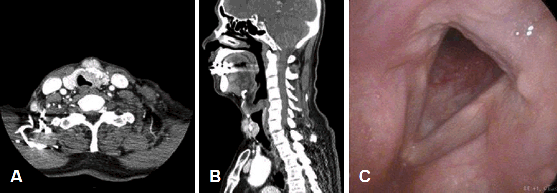
Preoperative neck CT. Axial and sagittal images of patient revealed a 3.4-cm low attenuated mass with calcification in the left thyroid gland (A and B). Subglottic invasion was shown by flexible endoscope (C).
The patient was treated with bilateral total thyroidectomy with central compartment neck dissection and tracheal window resection of the invading trachea. Both RLNs were preserved. There was an invading lesion overlaying the upper three tracheal rings. A tracheal defect measuring 20×35 mm was excised (Fig. 2). The defect was reconstructed with an ALT free flap. A vascularized ALT free flap (40×70 mm) was harvested from the left thigh (Fig. 3A). Arterial and venous anastomoses were performed to the superior thyroid artery and the branch of internal jugular vein, respectively (Fig. 3B). The fascial side of the ALT was sutured close to the tracheal defect using Vicryl®(Ethicon, Germany) 3-0 to maintain the appropriate position (Fig. 3C). Skin closure was then carried out in layers. To monitor the free flap, a small elliptical portion of the skin paddle of the ALT was designed and exteriorized between the upper and lower neck skin flaps (Fig. 3D). Official postoperative histopathology revealed PTC invading the tracheal cartilage and extending to the strap muscle. Central compartment lymph nodes were negative for malignancy. Endotracheal tube was removed about 12 hours after the end of the operation. The postoperative clinical course was uneventful and the patient was discharged on the postoperative day 18.
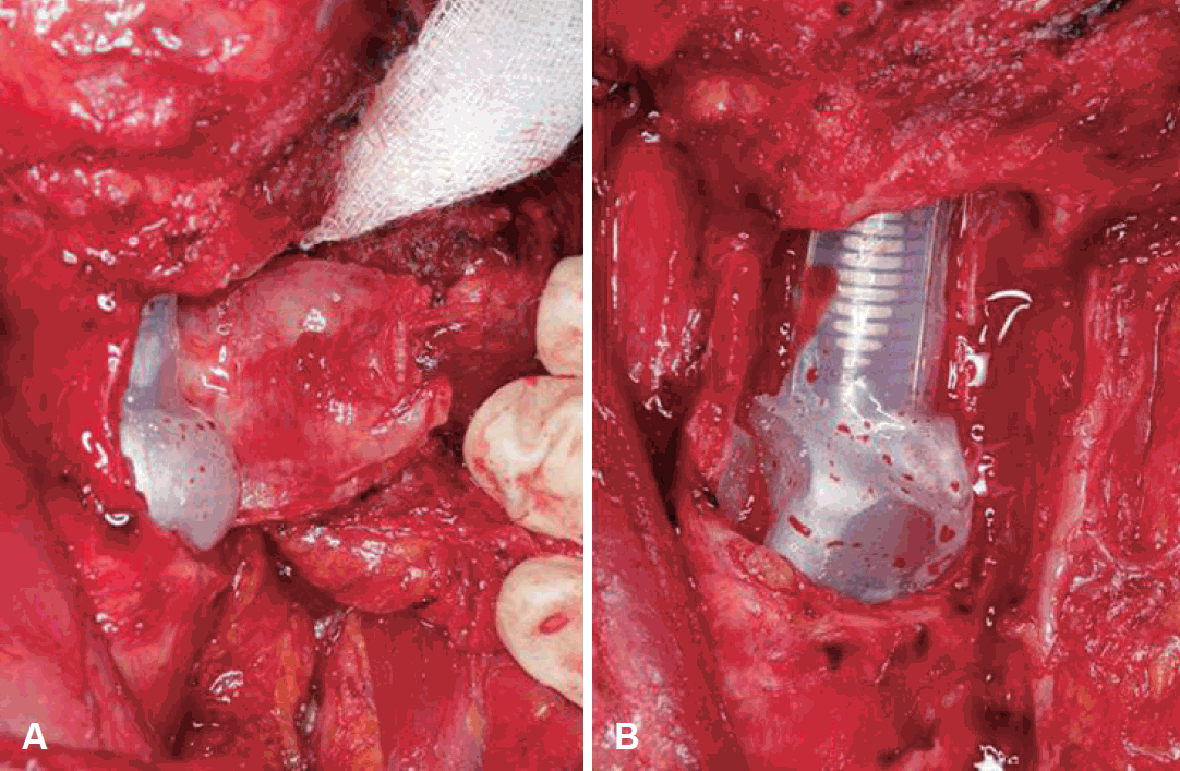
Tracheal defect measuring 20×35 mm (A). Tracheal defect after excision (B).
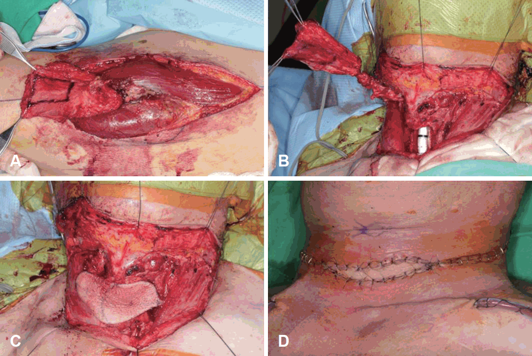
A vascularized anterolateral thigh free flap (40×70 mm) was harvested from the left thigh (A). Arterial and venous anastomosis were performed to the superior thyroid artery and the branch of internal jugular vein, respectively (B). Fascial side of the anterolateral thigh free flap was sutured close to the tracheal defect (C). The skin of anterolateral thigh free flap was designed and exteriorized between upper and lower neck skin flaps for the monitoring of the flap (D).
The patient underwent therapeutic high-dose (150 mCi) radioIodine-131 ablation at five months postoperatively. At 11 months postoperatively, follow-up CT scan of the patient’s neck and flexible endoscopy revealed no evidence of recurrence and normal vocal cord mobility (Fig. 4A, B, and C). And the flap site has engrafted in the recipient site (Fig. 4D). The patient’s thyroglobulin level was stable at less than 0.1 ng/mL. At the time of writing, 12 months had passed following surgery.
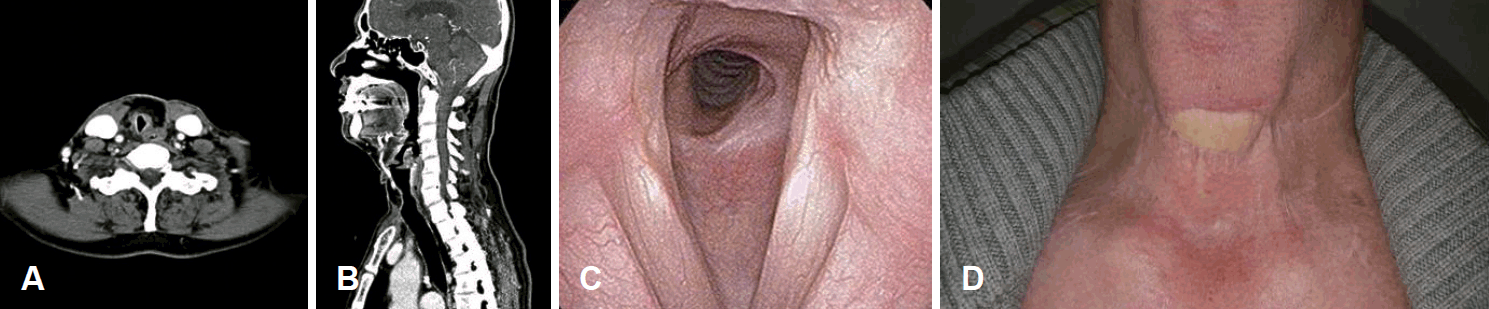
Follow-up CT scan of the patient’s neck revealed no evidence of recurrence (A and B). Follow-up flexible endoscopy revealed normal vocal cord mobility and patent airway in the subglottic area (C). Flap site engrafted in the recipient site at 11 months postoperatively (D).
Discussion
Organs other than the thyroid gland are susceptible to invasion in well-differentiated advanced thyroid cancer [2]. As we mentioned previously, these organs include the strap muscles, RLN, trachea, esophagus, and larynx. Local invasion often results in complications such as dyspnea, dysphagia, and voice change. Treatment involves en bloc surgical resection of the tumor with maximal preservation of function [6]. McCaffrey [7] proposed treatment options according to tracheal invasion stage (I-V) in patients with well-differentiated thyroid cancer. These stages include total thyroidectomy alone for stage I, total thyroidectomy and shave partial thickness excision of cartilage and/or muscle for stages II and III, and complete aerodigestive tract resection for stages IV and V.
Reconstruction might needed after tracheal resection. Secondary reconstruction by local flap [8], Pectoralis major myocutaneous flaps and free costal cartilage grafts [9], modified infrahyoid myocutaneous flaps [10], non-vascularized composite nasal septal grafts [11], latissimus dorsi musculocutaneous flaps [12], and free radial forearm flaps and titanium mesh [13] were reported for use in tracheal reconstruction. It is difficult to discuss the advantages and disadvantages of each method because there are no studies comparing cases and no longterm follow-up. In addition, some papers have mentioned the use of a radial forearm free flap with buccal mucosa in tracheal reconstruction [14,15] to prevent tracheal stenosis. In our case, fascia of the vastus lateralis muscle was used without a mucosal surface lining the trachea. Nevertheless, no problems occurred in the upper airway. This method also alleviated the need for buccal mucosa harvest.
Vascularized ALT is a fasciocutaneous flap including fascia of the vastus lateralis muscle. We expected the vastus lateralis muscle fascia facing the tracheal lumen to create a compatible airway. Tracheal resection and end-to-end anastomosis were also possible for our patient’s tracheal defect. However, side effects such as anastomotic leakage, rupture, and recurrent laryngeal nerve damage were possible. Reconstruction with a vascularized ALT flap such as the patch type can avoid these side effects, allowing operation with tracheal resection and end-to-end anastomosis when a vascularized ALT flap fails. In this case, successful treatment resulted from reconstruction using a vascularized ALT in well-differentiated advanced thyroid cancer invading the trachea. There was slight granulation tissue in the airway at the first postop OPD, but the airway was cleared in the final OPD follow-up. A buried flap is not easy to monitor, but it was possible by exposing the donor’s skin flap at the neck. Other drawbacks include unsatisfactory anterior neck appearan ce, longer operation time than with a local flap, and longer admission period. Verification of the stability and effectiveness of this surgical technique is needed and should be investigated using more cases with long-term follow-up.