접형동 점액낭종으로 인한 좌측 활차신경 단독 마비
Isolated Left Trochlear Nerve Palsy Caused by Sphenoid Sinus Mucocele
Article information
Trans Abstract
Paranasal sinus mucoceles are an uncommon cause of isolated palsies of cranial nerves III, IV, and VI. The trochlear nerve has been reported to be less frequently affected than the abducens and oculomotor nerves. Isolated sphenoid sinus diseases may cause serious complications by involving adjacent vital structures such as the optic nerve, cavernous sinus, internal carotid artery, and cranial nerves III–VI. We report a case of a 76-year-old woman who presented to our emergency department with a chief complaint of acute double vision and headache. Her diplopia was diagnosed as left trochlear nerve palsy. Brain CT and MRI revealed expanding cystic lesions in both sphenoid sinuses with bony erosion of the left sinus wall. The patient underwent an endoscopic intranasal sphenoidotomy and recovered completely from diplopia at postoperative 2 months. The relationship between the trochlear nerve palsy and its anatomy is also discussed.
서 론
외안근마비는 복시와 시력장애를 일으키는 질환으로 뇌신경 3, 4, 6번 마비에 의해 주로 발생한다. 단독 마비는 대부분 3번과 6번이고 4번은 드물며 복합 마비는 3번과 6번, 또는 세신경 모두의 조합이 많다. 급성 마비의 주된 원인으로 동안 신경은 혈관병 및 동맥류, 외전신경은 다발성경화증이나 수막뇌염 같은 염증성 질환, 그리고 활차신경은 머리 외상에 의해 가장 많이 발생한다고 보고되었다[1]. Mollan 등[2]은 성인의 상사근(superior oblique muscle) 마비 원인을 선천성, 외상, 혈관병의 순으로 보고하였다. 비외상성 후천성 활차신경마비는 당뇨, 고혈압, 고콜레스테롤혈증 등이 동반될 때 미세혈관성으로 분류되고, 혈관위험인자가 없을 때 특발성으로 분류되는데 이들은 뇌영상에서 두개 내 병변이 발견되지 않고 보통 수주에서 수개월 내에 자연 회복되는 경우가 많은 것이 특징이다[2,3]. 그러나, 뇌신경 3, 4, 6번 마비와 연관된 부비동질환은 매우 드물어 원인의 한 범주를 차지하고 있지 않다. 접형동의 단독 병변은 모든 부비동 병변 중에서 3% 이내로 알려져 있고 가장 흔한 원인 인자는 염증성 병변으로 10~17%를 점액낭종이 차지한다[4]. 부비동 점액낭종은 부비동 내에 점액이 저류되는 확장성 낭종성 병변으로, 특히 접형동은 시신경, 해면정맥동, 내경동맥, 그리고 3, 4, 6번 뇌신경 등과 인접해 있어 팽창하는 낭종이 이들 중요 구조물과의 경계가 되는 얇은 골판을 쉽게 침범하여 임상 증상을 일으키게 된다[5]. 그러므로 접형동 점액낭종은 양성 질환이지만 시신경관(optic canal)이나 상안와열(superior orbital fissure)을 침해했을 때는 시력 상실이나 복시 등의 시각장애를 초래하는 임상적으로 중대한 질환이 될 수 있다. 접형동 단독 병변에서 시신경이 가장 영향을 많이 받고 그 다음으로는 보고자에 따라 동안신경이나 외전신경이라고 알려져 있다[6,7]. 그러나 활차신경마비는 매우 드물어 사골동과 접형동 점액낭종에서 시신경병(optic neuropathy)과 동반된 1예가 보고되었을 뿐이다[8]. 국내에서도 접형동 점액낭종에 의한 시신경병과 동안신경 및 외전신경 단독 마비는 각각 보고되었으나[9-11] 활차신경 단독 마비는 아직까지 보고된 예가 없다. 저자들은 최근 접형동 점액낭종에 의한 활차신경 단독 마비를 접형동 수술로 치료한 경험을 활차신경의 해부학적 고찰과 함께 보고하고자 한다.
증 례
76세 여자 환자가 2일 전 발생한 좌측 두통과 복시로 응급실에 왔다. 2주 전부터 좌측 측두두정 부위에 시각통증등급 6~7점가량의 압박성, 박동성 통증이 간헐적으로 반복되었고 강도는 점점 심해지는 양상이었다. 2일 전부터는 수직 복시가 발생하여 병원에 왔고 환자는 한쪽 눈을 가렸을 때 복시가 사라졌으며 단안시에서는 시력 저하를 느끼지 않는다고 말하였다. 고혈압으로 약을 복용 중이고 25년 전 머리 외상으로 후두부에 인공두개골 삽입술을 받은 병력이 있었다. 사시 검사에서 안구는 제1안위에서 경도의 좌측 상사시(hypertropia)를 보였고 수직 편위도가 좌측 상사근의 작용방향인 우하방 주시, 즉 좌안 내회선(intorsion) 때에 커져 좌측 상사근마비로 판별되었다(Fig. 1). 시력을 포함한 다른 신경학적 검사에서는 이상 소견을 보이지 않았다. 따라서 환자는 상사근을 지배하는 활차신경 단독 마비로 진단되어 원인 질환을 감별하기 위한 뇌영상검사를 시행하였다. 조영 증강 뇌 CT 및 MRI 축영상에서 양측 접형동을 채우고 있는 4.0×3.1 cm의 점액낭종에 합당한 소견이 확인되었고 좌측 안와첨(orbital apex)과 경사대(clivus)의 침범이 의심되는 소견을 보였다(Fig. 2). MRI 관상영상에서는 해면정맥동의 압축이 관찰되었다(Fig. 3A). 비내시경검사에서 비강 내 이상 소견은 관찰되지 않았다. 복시 발생 4일째 전신마취하에서 내시경 비강 내 접형동절개술을 시행하였다(Kim YH). 우측 접형동에서는 고름이 배농되었고 좌측 접형동에서는 단순 점액을 지닌 점액낭종이 확인되었다. 낭종벽을 제거하고 배액 후 부비동을 세척하였으며 접형동 내로 중요 구조물들의 노출은 보이지 않았다. 양측 접형동 자연공을 전하방으로 넓게 열어 주고 양측 접형동 사이 중격을 제거한 후 합병증 없이 수술을 마쳤다. 세균배양검사에서 포도상구균이 동정되었고 입원기간 동안 항생제로 cefotetan disodium(2 g/day)을 사용하였으며 전신 스테로이드는 투여하지 않았다. 환자는 수술 직후 두통은 호전되었고 복시 증상은 남아 있었으나 이것도 점차 호전되어 수술 2개월째에 완전히 소실되었고 육안으로 보아 상사근 마비도 관찰되지 않았다. 수술 4개월째에 시행한 부비동 CT에서 좌측 접형동 후벽의 점막 비후 이외에 연조직 음영은 관찰되지 않았고(Fig. 3B) 환자도 특별히 호소하는 증상 없이 현재까지 추적 관찰 중이다.

Left trochlear nerve palsy in the patient with left sphenoid sinus mucocele. In primary gaze, mild incomitant hypertropia and excycloduction of the left eye are findings compatible with left superior oblique palsy (A). In right gaze, hypertropia and loss of intorsion is observed on the left eye (B). In left gaze, normal conjugate eye movement is demonstrated (C).

Mucocele of left sphenoid sinus in axial contrast-enhanced CT scans (A and C) and T2-weighted MRI (B and D). CT scans demonstrate nonenhancing opacification of both sphenoid sinuses, which is T2 hyperintense in the MRI. The heterogeneous intensity in the MRI may be related with the mucin concentration of the mucocele. The orbital apex (arrows) and clivus (arrowheads) are involved with bony erosion.
고 찰
부비동 점액낭종의 발병기전은 명확하게 밝혀져 있지 않지만 자연공의 만성 폐쇄로 인해 점액 배출 장애가 발생되고 이로 인해 점점 커지는 낭종을 형성한다고 알려져 있다[12]. 접형동 질환의 가장 흔한 발현 증상은 두통으로 33~81%의 발병률을 보이고 두개안면부 어느 부위에서나 나타날 수 있다. 두 번째로 흔한 발현 증상은 시각장애로 단독 접형동 질환의 24~50%에서 나타난다고 보고되었다[13]. 점액낭종이 시각장애를 유발하는 기전은 인접한 염증에 의한 신경염, 팽창하는 병변의 압력에 의한 허혈, 그리고 혈전정맥염/혈관염에 의한 경색으로 설명할 수 있다[7]. 4번 뇌신경인 활차신경은 뇌신경 중에서 두개 내 경로가 가장 길고 두께가 가장 얇고 작은 신경이다[14]. 활차신경핵은 하구(inferior colliculus) 위치의 중심선 근처 복측 회백질에 위치해 있는데 활차신경은 배측 중뇌에서 단일 뿌리로 시작되어 하구의 하단 외측으로 나온다. 신경은 상소뇌각 외측을 지나 교뇌 바로 위 대뇌각을 감돌아 소뇌천막(tentorium cerebelli) 밑에서 주행한 후 추체침대 주름(petroclinoid fold) 아래에서 경사대 측면을 따라 경막을 관통하여 해면정맥동의 외측벽을 뚫고 안으로 들어가 천장 아래 측벽에서 동안신경 아래에 놓여 정맥동을 통과한다. 상안와열(superior orbital fissure)은 외직근에 의해 상외측부와 하내측부로 나뉘는데 활차신경은 상외측부를 통해 안구 내로 진입하여 안와지방 내에서 내측 대각선 방향으로 돌아 상사근에 도달한다[2,14].
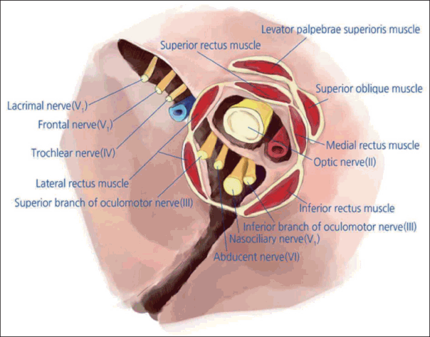
본 환자에서 활차신경 단독 마비가 일어난 기전은 접형동 외벽의 침식으로 인한 낭종의 해면정맥동 압박보다는 안와첨(orbital apex)에서 활차신경의 해부학적 위치와 관계가 있을 것으로 추정된다. 활차신경은 해면정맥동 내에서 측벽의 위쪽 가장자리 동안신경의 1 mm 아래에 위치하기 때문에[14] 접형동 병변이 해면정맥동으로 침범했다면 다발성 뇌신경마비와 동반되었을 것이다. 안와 내에서 활차신경은 뇌신경 3, 4, 6번 중 유일하게 네개의 직근이 기원하는 총건륜(annulus of Zinn, common annular tendon) 바깥쪽에 위치하여 주행한다(Fig. 4) [14]. 이러한 특징적 경로가 신경 손상에 대한 취약성에 기여할 것으로 생각되는데, CT와 MRI에서 보이는 골침식으로 점액낭종이 안와첨을 침범하였다면 총건륜 밖에 노출되어 있는 활차신경에 염증이 침습하여 단독 마비가 올 수 있다고 생각된다. 접형동 점액낭종의 치료는 수술적으로 낭종을 제거하고 점액 배출이 용이하도록 접형동의 전하벽을 넓게 열어주는 것인데 최근 내시경 부비동 수술의 발달로 고식적인 외비접근법은 대부분 내시경 수술로 대체되었다[15]. Lee 등[7]은 시각장애를 보이는 접형동 단독 질환 환자들에서 시력 저하는 내시경 수술 후에도 비가역적인 경우가 많으나 복시는 높은 호전율을 보인다고 하였으므로 본 증례와 같은 복시 환자에서 신속한 진단과 수술의 중요성이 강조된다.

Schematic drawing of the superior orbital fissure with its components at the right orbital apex. The trochlear nerve emerges in the lateral part of the superior orbital fissure, just lateral to the annulus of Zinn from which the four rectus muscles origin. The course of the trochlear nerve outside the muscle cone may contribute to its vulnerability to injuries.
저자들은 복시와 두통을 호소하는 환자에서 활차신경 단독 마비와 그 원인이 된 접형동 점액낭종을 진단하고 비강내 접형동절개술로 이를 치료하였기에 발병 기전의 해부학적 고찰과 함께 보고한다.
Acknowledgements
We thank Bora Crystal Seo for the medical illustration.
