측경부에 발생한 혈관 내 유두양 혈관내피세포 증식증 1예
A Case of Masson’s Tumor in Lateral Neck
Article information
Trans Abstract
Masson’s tumor, also known as intravascular papillary endothelial hyperplasia (IPEH), is a rare, benign vascular tumor characterized by the proliferation of endothelial cells with papillary formations. Differential diagnosis between IPEH and angiosarcoma is important because both have microscopic similarity. Herein, we report a rare case of IPEH on the right lateral neck of a 50-year-old female presenting with a neck mass, which was completely removed without complication.
서 론
혈관 내 유두양 혈관내피세포 증식증(intravascular papillary endothelial hyperplasia, IPEH)은 피부와 연조직에 발생하는 혈관 종양 중 2%에 해당하는 드문 질환으로 50세 이하의 젊은 환자에서 발생하는 경우가 많고, 여성에서 경미하게 발생 빈도의 우위를 보이며, 전신 어디에나 발생할 수 있으나 측경부에서 발견되는 것은 매우 드문 경우이다[1,2]. IPEH는 혈관내피세포의 과도한 유두양 증식을 특징으로 하는 혈관 내 양성 반응성 병변으로 임상적으로나 병리학적으로 혈관 육종과 아주 유사하기 때문에 이를 정확하게 감별하는 것이 치료에 매우 중요하다[3,4].
저자들은 우측 경부 종물을 주소로 내원하여 측경부의 IPEH로 진단받은 환자를 경험하여 문헌고찰과 함께 보고하는 바이다.
증 례
50세 여자 환자가 5개월 전부터 촉지되는 통증 없는 우측 경부 종물을 주소로 내원하였다. 발열, 호흡곤란, 연하곤란 등의 증상은 없었다. 병력 청취상 수술받은 병력 및 최근에 외상을 입은 적은 없었고 좌측 양성 갑상선 결절 외 진단받은 기저 질환은 없었다.
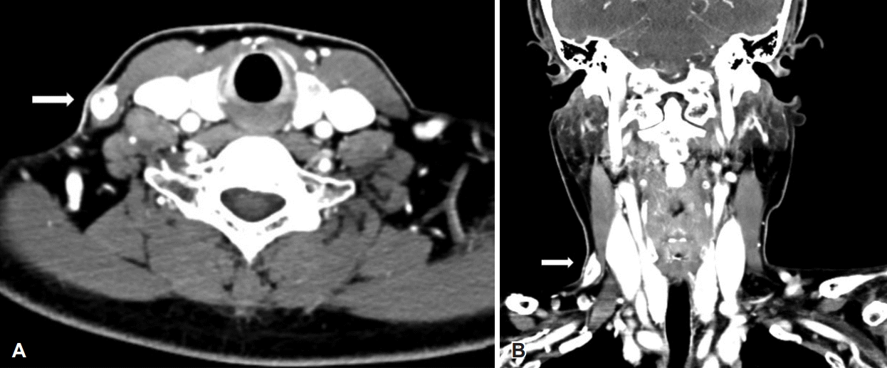
신체검진상 우측 경부 피하의 10×10 mm 크기의 무통성의 종물이 촉지되었고 그 외 다른 경부 종물은 없었다(Fig. 1). 경부 초음파와 조영 증강된 경부 컴퓨터단층촬영(enhanced neck CT)을 시행하였다. 경부 초음파상, 우측 경부 구역 Vb에 11×5×15 mm 크기의 타원형의 강한 에코를 보이는 종물이 확인되었다. Neck CT상, 우측 흉쇄유돌근 위에 위치하는 타원형의 조영 증강되는 10×19 mm 크기의 종물이 우측 전경정맥으로 배액 되는 말초혈관과 연결된 것을 확인하였다(Fig. 2).

Preoperative findings of enhanced CT scan. (A) is axial and (B) is coronal view showing oval shaped mass, measuring 10×19 mm, situated over the right sternocleidomastoid muscle, with a peripheral vessel draining into the right anterior jugular vein (arrow).
이에 조직학적 진단을 위해 전신마취하 우측 측경부 종물 절제술을 시행하였고, 우측 흉쇄유돌근 위에 위치한 10×10×5 mm 크기의, 혈관이 관통하고 있는 종물을 완전 절제하였으며 수술 중 출혈은 많지 않았다.
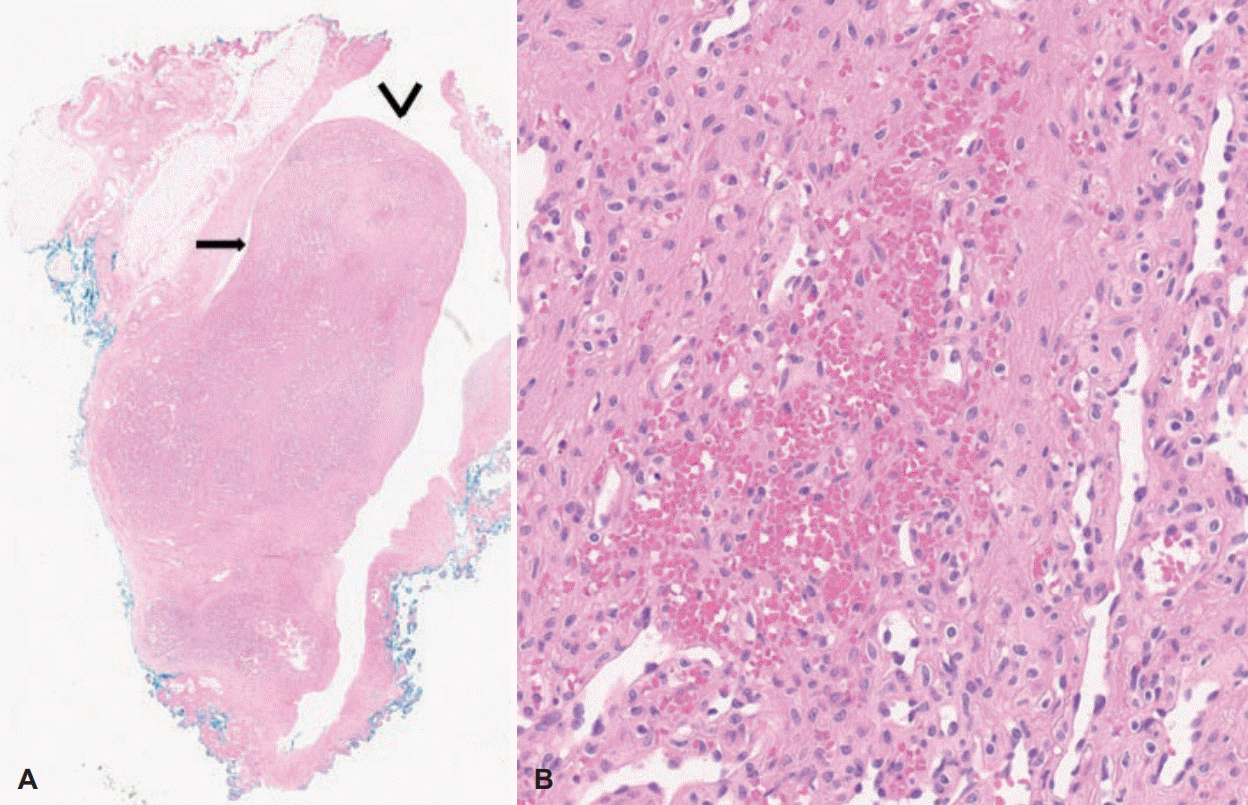
절제된 조직은 병리 소견상 확장된 혈관벽에서 생긴 결절성 병변으로, 저배율에서 유두양으로 성장하는 패턴을 보이는 방추세포로 이루어진 중간엽 종양으로 생각되었다. 고배율에서 이 방추세포들은 핵의 비정형성을 보이지 않아 양성의 혈관 내피 세포로 사료되어 IPEH로 진단하였다. 괴사 세포 분열 혹은 핵의 비정형성은 보이지 않아 혈관 육종과 감별할 수 있었다(Fig. 3). 수술 다음날 특이 합병증 없이 퇴원하였고, 1주 뒤 외래 종결하였다.

Microscopic finding of intravascular papillary endothelial hyperplasia. (A) is showing proliferation of papillary structures (arrow) within intravascular space (black v shape, H&E stain ×10). (B) is showing no cellular atypia or necrosis in intravascular papillary endothelial hyperplasia (H&E stain ×200). H&E: hematoxylin and eosin.
고 찰
IPEH는 1923년 프랑스 병리학자 Masson [3]에 의해 처음으로 언급된 혈관 육종과 구별되는 혈관의 양성 과형성 질환으로 Masson’s tumor, Masson’s hemangioma 등으로 다양하게 일컬어져왔다. 1976년 Clearkin과 Enzinger [5]가 소개한 IPEH라는 용어가 현재 가장 널리 사용되고 있다. IPEH는 전신 어디에나 발생하는 것으로 알려져 있으며 주로 상하지, 두경부 부위에서 발생하는 것으로 알려져 있으나 측경부에서 발견되는 경우는 매우 드물다. 문헌 고찰상 본 증례와 같이 측경부에서 발생한 증례는 국외에서 3예가 보고되었으나 국내에서는 보고되지 않았다(Table 1) [6-8].
병인에 대해서는 명확히 밝혀져 있지 않으나 Salyer와 Salyer [9]는 IPEH에서 혈전의 조직화 과정에서 보이는 특징적인 형태가 관찰된다고 기술하여 혈전 생성이 원인일 수 있다는 견해가 있다. Hashimoto 등[4]은 IPEH를 본 증례와 같이 확장된 혈관에서 발생하는 원발형(56%), 화농성 육아종에서 발생하는 혼합형(40%), 그리고 혈종에서 발생하는 경우인 혈관외형(4%)의 3가지 형태로 구분하였다.
진단을 위해 육안적으로 검진한 후 병변의 성상과 크기를 확인하기 위해 영상학적 검사로 neck CT를 촬영할 수 있다. 이 경우 균질한 조영 증강, 불균질한 조영 증강을 나타내는 경우[3,10] 등 병변이 다양하게 관찰될 수 있으며 본 증례에서는 병변이 경부에 국한된 균질한 조영 증강 소견을 보였다. 또한 자기공명영상도 촬영할 수 있는데 T1 강조영상에서 중간신호 강도를, T2 강조영상에서 고신호강도를 보인다[11].
이와 같이 영상학적 검사만으로는 IPEH를 혈관종, 혈관 육종, 악성 종양과 감별하기 어려워[12] 확진을 위해서는 조직 검사를 통해 특징적인 조직학적 소견의 확인이 필수적이다. 병리학적으로 IPEH는 혈관 내로 혈관 내피 세포의 유두양 팽창을 보이는데 이는 혈관종 및 혈관 육종에서도 나타나는 특징이므로 다른 질환들과 감별하는 것이 중요하다. IPEH에서는 혈관 내피 세포의 팽창된 유두의 개수가 많고 조밀하게 배열되는 반면 혈관종에서는 선형으로 배열된다[13]. 혈관 육종과 구분되는 IPEH의 특징은 주변 조직의 침범 없이 혈관 내에 병변이 국한되며 많은 수에서 혈전이 동반되고 세포의 비정형성 및 내부에 괴사조직이 보이지 않는다는 점이다[1,6].
본 증례의 경우 내피 세포의 증식이 혈관 내에서만 관찰되며 종괴 내부에 비정형 소견 및 괴사 조직이 관찰되지 않아 혈관 육종을 배제하고 IPEH로 진단하였다.
IPEH의 치료는 외과적 절제이며[6] 뇌나 두개골 등 위험한 부위의 수술 시에는 혈관 조영술 혹은 수술 전 색전술을 시행할 수 있으나, 모든 부위에 대해서 시행하지는 않는다[14]. 재발률은 낮으나 병변이 완전히 제거되지 않았을 경우 재발할 수 있고 재발한 경우에도 병변의 완전 절제를 목적으로 치료를 시행한다. 이와 달리 혈관 육종은 광범위한 수술적 절제 및 술후 방사선 치료를 요하는 경우가 많고 국소 재발 및 원격 전이가 흔히 발생하며 5년 생존률이 10~20%라고 보고된 예후가 불량한 질환이기 때문에, 적절한 치료를 위해 IPEH와 혈관 육종을 정확히 감별하는 것이 중요하다[15].
본 증례는 측경부에 발생한 IPEH를 합병증 없이 제거한 증례로 경부의 조영 증강이 되는 종괴가 있을 때 감별해야 하는 질환들 중 드물지만 IPEH를 염두에 두어야함을 상기시키는 증례이다. 또한 IPEH는 혈관 육종과 유사한 특성을 가지나 조직학적으로 구분되는 점을 잘 기억하여 정확한 진단을 내리고 단순 절제를 통해 치료하는 것이 중요하다.
Acknowledgements
None.
Notes
Author Contribution
Conceptualization: Hoyoung Lee, Soo Jeong Choi. Data curation: Hoyoung Lee, In Hak Choi. Supervision: Kwang Yoon Jung. Writing—original draft: Hoyoung Lee. Writing—review & editing: Hoyoung Lee, Kwang Yoon Jung.


