내시경 고실성형술의 다양한 술식
Variations of the Technique in Endoscopic Tympanoplasty
Article information
Trans Abstract
Endoscopic tympanoplasty is a surgical procedure for patients with tympanic membrane perforation with minimal middle ear or mastoid inflammation. Recent findings revealed that endoscopic tympanoplasty harvests equivalent or even superior results over microscopic tympanoplasty. However, a number of disadvantages are related to endoscpoic tympanoplasty, one of which is the single-handed procedure that may lead to recurrent perforation. We hereby illustrate a number of techniques involved in endoscopic tympanoplasty along with their pros and cons.
서 론
고실성형술은 중이강이나 유양돌기에 염증이 심하지 않은 고막천공의 경우 많이 사용되는 방법이다[1]. 최근 내시경을 이용한 고실성형술이 보고되었고 이 술식의 결과가 현미경을 사용한 경우보다 좋거나 동등한 결과들을 보이고 있다[2,3]. 또한 수술 중 불편감이나 술후 통증이 현미경적인 수술보다 좋다는 보고가 있어. 앞으로 더 많은 환자들에게 적용될 가능성이 있다[2].
그러나 한 손으로 내시경을 잡고 다른 한 손으로 술식을 진행하기 때문에 연골막이나 근막의 삽입이 용이하지 않은 경우가 있어서 술후 재천공이 발생하는 경우도 보고되고 있다[4,5]. 이런 재천공은 주로 천공의 앞쪽 부분에 발생하는데 이는 연골막을 삽입하고 젤폼을 이용한 지지가 충분치 못해 발생하는 것으로 생각된다.
이에 따라 저자들은 외이도를 통한 내시경적 고실성형술의 술식을 두 가지 다른 방법으로 소개하고자 한다.
방 법
천공변연부 합입된 피부조직의 제거
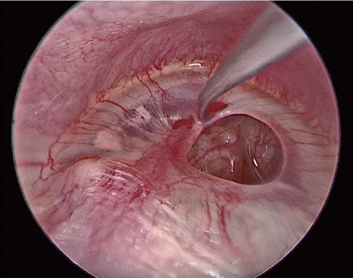
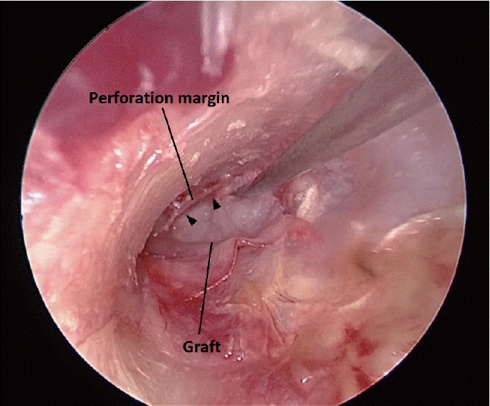
고막천공의 경우 천공된 변연부를 따라 상피세포가 자라서 안쪽으로 들어가 있으므로 변연부를 충분히 제거하여 고막상피세포가 재생할 수 있도록 만들어준다(Fig. 1).
이주 연골의 연골막 채취
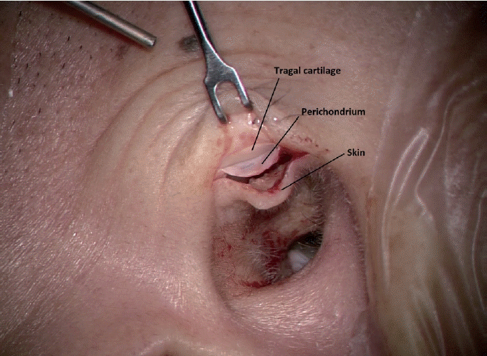
내시경적 고실성형술의 경우 후이개절개를 하지 않으므로 이주부위를 절개하여 연골막을 채취한다. 이때 미리 천공의 크기를 측정하여 충분한 크기의 연골막을 채취하도록 한다(Fig. 2).
고실 내 관찰
고실 내의 이소골과 중이점막 상태를 관찰한다. 이소골은 추골병과 침등관절을 관찰하여 이소골 연쇄가 손상되지 않았는지 확인한다.
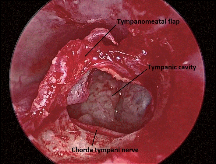
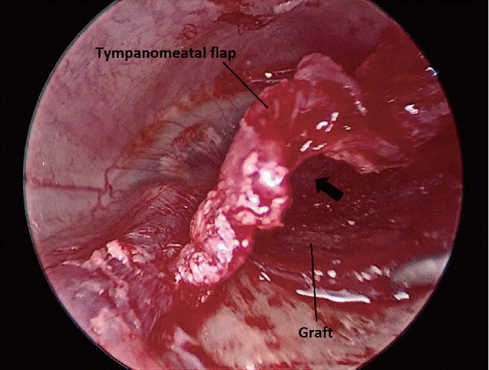
외이도 피부에 절개선을 넣어 고막을 앞쪽으로 거상하는 transcanal 방법의 경우는 보다 넓은 부위를 관찰할 수 있으며 이 경우 고삭신경이 손상되지 않도록 주의하여야 한다(Fig. 3).
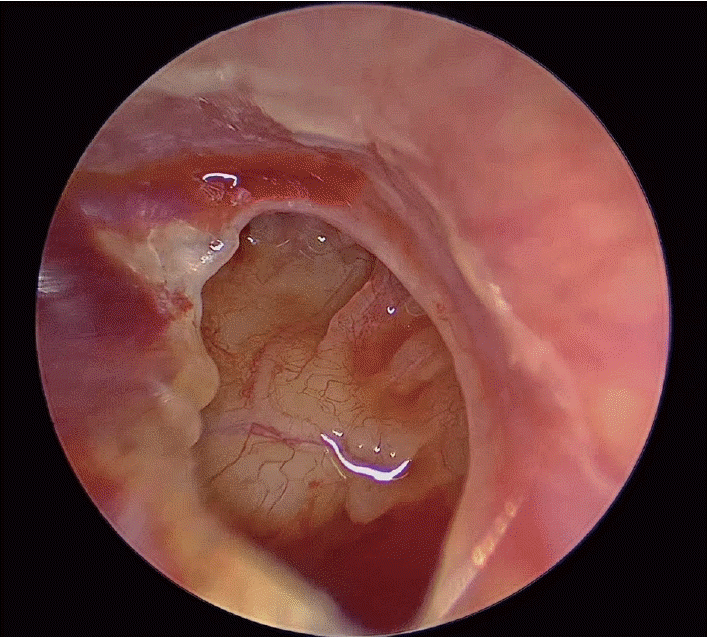
Endocanal 방법은 천공부위를 통해서 고실 내를 관찰하며 0도 내시경 또는 30도 내시경을 이용하여 관찰한다(Fig. 4).
연골막의 삽입
연골막의 삽입 방법은 기본적으로 두가지로 나누어진다. Endocanal 접근법을 이용하는 경우는 연골막을 천공부위를 통해서 넣는 push through 방법을 이용한다. 천공부위를 통해 젤폼을 미리 고실 내에 채우고 연골막을 천공부위에 삽입하여 고막의 내측으로 넣는다(Fig. 5).
Endocanal 접근법은 기존의 microscopic myringoplasty와 같은 개념의 술식으로, anterior perforation의 위험이 적은 장점이 있는 반면, 이소골 연쇄와 중이 내부 상태를 탐색하기에 상당한 제한이 따르는 단점이 있다.
Transcanal 접근법의 경우는 거상된 고막부위 아래로 뒷쪽에서부터 연골막을 삽입하고 젤폼을 이용하여 고실의 앞쪽부터 채워서 연골막을 고막쪽에 지지시킨다(Fig. 6).
Transcanal 접근법의 경우 앞쪽의 천공부위에 삽입된 연골막이 젤폼으로 충분히 지지되지 못하면 재천공이 발생하므로 앞쪽의 연골막을 들고 추가로 젤폼을 채워서 연골막이 고실쪽에서 충분히 지지될수 있도록 한다.
Transcanal 접근법은 endocanal 접근법과 달리 anterior perforation의 위험성이 높다는 단점이 있는 반면, 이소골 연쇄와 중이 내부 상태를 탐색하는 것이 용이하다는 장점이 있다.
이 두 가지를 합하여 사용하는 combination technique을 사용할수도 있다. 이는 push through 방법으로 연골막을 삽입하고 transcanal 접근법에서와 같이 고막을 거상하여 뒷쪽으로 들어간 연골막을 고막내측으로 위치시키는 술식이다(Supplementary Video 1).
고막외측 부위의 처치
연골막을 삽입한 후 고막의 외측은 젤폼 등으로 채워서 삽입된 연골막이 움직이지 않도록 한다.
결 과
내시경적 고실성형술은 현미경적 고실성형술과 비교했을 때 몇 가지 차별점이 있는 것으로 알려져 있다. One-hand technique임에 따라 지혈 등 수술이 다소 까다로울 수 있으며 내시경으로 인한 thermal damage의 가능성이 있을 수 있는 반면, 현미경에 비해 수술적 시야 확보가 용이하여 canalplasty가 필요한 경우가 드물어 덜 침습적이며 수술 시간도 더 짧은 것으로 보고되고 있다[6]. 이러한 내시경적 고실성형술은 endocanal 접근법이나 transcanal 접근법이 모두 유용하게 사용될 수 있다. 고막천공의 위치, 이소골 상태, 이소골 주위 중이점막의 상태 등을 고려하여 적절한 술식을 사용하면 재천공의 가능성을 줄이고 통증을 줄이는 방법으로 수술을 마칠 수 있다.
Supplementary Material
The Data Supplement is available with this article at https://doi.org/10.3342/kjorl-hns.2021.00493.

