외비에서 발생한 선암종 1예
A Case of Adenocarcinoma of the External Nose
Article information
Trans Abstract
Adenocarcinoma occurs very rarely in the paranasal sinuses; moreover, adenocarcinoma diagnosed in the external nose is rarely reported. A 58-year-old male patient visited our hospital with an enlarged right nasal mass. On image studies, a 1.5 cm sized mass was found on the right nasolabial fold, which was surgically removed via sublabial approach. Intraoperative frozen section examination showed inflammatory tissue without tumor cells. However, adenocarcinoma showing infiltration between the skeletal muscle layers was diagnosed on the final histopathologic examination after surgery. In the immunohistochemical staining test, CK7 positive, CK20 negative, and GCDFP-15 positive were confirmed, so metastasis of adenocarcinoma was considered first rather than primary cancer of the nasolabial fold. There was no recurrence and no tumor development in other sites after postoperative chemoradiation therapy of 32 months.
서 론
선암종(adenocarcinoma)은 보통 소화기계, 호흡기계, 유방, 전립선, 부인과계에서 주로 발생하는 악성종양이며[1], 비부비동으로 전이되어 진단되는 경우는 드물게 보고되고 있으나 원발부위를 찾을 수 없이 외비(external nose)에서 발견된 사례는 보고된 바 없다. 비부비동의 원발 암종은 많은 경우에서 진단되고 치료가 진행되고 있으나, 비부비동으로의 전이암은 상대적으로 매우 드물다[2]. 전 세계적으로 비부비동으로의 전이암은 1951년부터 2011년까지 총 167건이 보고되어 있고, 그중 신세포암에서 가장 많은 비부비동 전이를 보였으며 전이 위치는 상악동으로의 전이가 가장 많았다[2]. 선암종의 비부비동으로의 전이는 Batson [3]에 의해 추골정맥총을 통한 전이 과정이 설명된 적 있다. 본 증례와 같이 외비(external nose)에서 원발 선암종이 확인되거나 선암종이 전이된 경우는 매우 드물어서, 저자들은 58세 남성 환자의 우측 외비에서 확진된 선암종 1예를 재발 소견 없이 성공적으로 치료하여 문헌 고찰과 함께 보고하고자 한다.
증 례
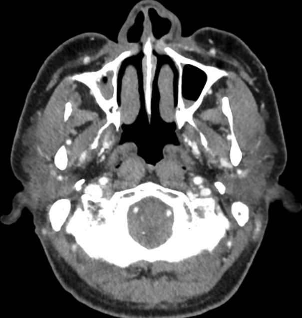
58세 남자 환자가 1년 전부터 발견된 우측 비익과 코입술주름 사이 부위의 종물이 크기가 커지는 양상을 보여 이비인후과의원에서 본원으로 의뢰되었다. 과거에 외상력 및 코 관련 질환, 코 수술을 받은 적은 없었고, 비루, 비출혈 등의 증상은 없었으나 우측 코 안의 답답함을 호소하였다. 비내시경 검사에서 우측 비전정이 쳐져 있어 우측 비강 입구가 상대적으로 좁은 소견과 우측으로 비중격 만곡이 관찰되었다. 외형상 종물이 돌출된 양상은 아니었으나, 촉진 시 우측 코입술주름 부근에서 무통성의 종괴가 촉지되었다. 종물의 정확한 위치와 경계를 파악하기 위하여 부비동 전산화단층촬영, 자기공명영상을 촬영하였으며, 부비동 전산화단층촬영에서 우측 코입술주름에 1.5 cm 크기의 조영 증강되는 종물이 확인되었고, 자기공명영상 촬영에서 우측 코입술주름에 T2 강조영상에서 저신호 강도를 보이며 경계가 불명확한 종물이 관찰되었다(Fig. 1). 이런 소견을 종합하여 우측 코입술주름 부위의 육아종성 종물을 의심하였고, 정확한 조직학적 진단 및 제거를 목적으로 구순하 절개를 통한 종물 절제술을 시행하였다.
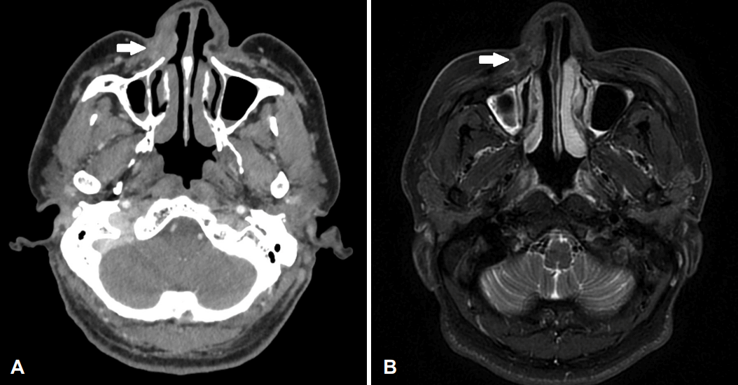
Contrast enhanced inhomogenous mass (arrow) in the right external nose (A, axial CT image; B, axial T2 MRI image).
수술은 비중격 교정술을 먼저 시행하였고, 그 후 우측 구순하 절개를 가한 뒤 상악골을 따라서 박리해 나갔다. 수술 중 우측 콧주름 하부 조직이 전반적으로 단단한 상태였고, 일부 제거하여 동결절편검사를 시행한 결과 종양 세포는 존재하지 않는 염증성 조직의 소견이 확인되었다. 주변 조직을 추가적으로 제거하여 조직검사를 시행하였으며, 주변부를 Coblator II controller (Arthrocare, Sunnyvale, CA, USA)로 제거 후 수술을 종료하였다. 환자는 특별한 합병증 없이 수술 2일 후 퇴원하였고, 수술 1주 후 외래 내원 시 비중격은 잘 교정되어 있었다. 입술 아랫부분의 절개 및 수술 부위는 잘 회복되고 있었으나, 우측 콧주름 부위의 지연성 화상 소견이 확인되어 연고 도포와 습윤성 드레싱을 시행하였다. 최종 조직검사 결과 골격근층 사이로 침윤성을 띄는 고등급 선암종으로 진단되었고(Figs. 2 and 3), 면역조직화학염색에서는 CK7 양성 및 CK20 음성, GCDFP-15 양성, TTF-1 음성, CK 양성이 확인되었다. 수술 후 시행한 전신 양전자방출단층촬영 검사상에서 다른 원발 부위의 이상 소견이나 전이 소견은 확인되지 않았다. 술전 영상 검사나 임상 양상 등에서 악성종양이 의심되지 않았고, 수술 중 동결절편검사에서 종양의 소견이 확인되지 않아 수술 시 충분한 절제 변연을 확보하지 못했기 때문에 조직검사 결과 확인 후 충분한 절제 변연 확보를 위한 추가적인 광범위 절제술을 고려하였다. 하지만 환자가 수술 후 흉터와 코 주변부 결손에 대한 우려로 거부하여 6600 cGy의 방사선치료와 동시에 cisplatin을 이용한 항암치료를 3차례 시행하였고, 수술 후 32개월 동안 추적 관찰 동안 재발이나 다른 원발 부위에 암종 소견은 관찰되지 않았다(Fig. 4).
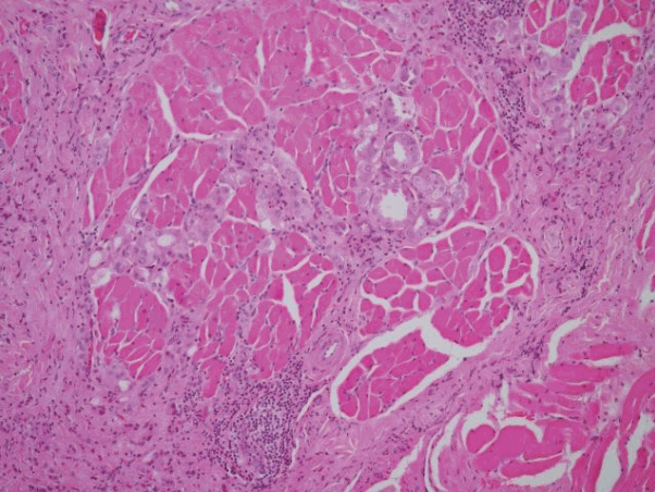
Findings of invasion of tumor cell nests that form part of the glandular structure between the skeletal muscle bundles (hematoxylin and eosin stain, ×100)
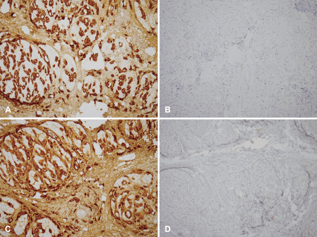
Immunohistochemical findings (×100). A: Positive for CK7. B: Negative for CK20. C: Positive for GCDFP-15. D: Negative for p63.
고 찰
일반적으로 코와 비부비동에는 편평상피세포암종, 타액선형 종양, 미분화암종, 흑생종, 후각신경모세포종, 신경내분비암종, 연부조직 종양 등의 악성종양이 발생한다[4]. 하지만 원발 부위를 찾을 수 없는 외비 선암종은 아직 보고된 바가 없다. 코에서 진단된 선암종은 원발암 또는 전이암을 고려할 수 있는데, 본 증례에서는 영상 검사에서 다른 원발 부위를 찾을 수 없었지만 면역조직화학염색상 CK7 양성 및 CK20 음성, GCDFP-15 양성을 보인 점을 고려하여 원발암보다 전이암의 경우를 더 의심하였다.
코에서 진단되는 선암종의 발병 요인에 대한 연구는 명확하게 밝혀진 바 없으나, 비부비동 선암종의 경우는 목재 분진, 포름알데히드, 금속 노출(니켈, 크롬), 탄닌(tannin) 공정, 만성 점막 염증 등이 발병에 영향을 주는 요인으로 고려되고 있다[5,6]. 비부비동 내 병변일 경우 편측 코막힘, 비루, 빈번한 비출혈 등의 증상이 발생 가능하나[7], 외비에 발생한 병변의 경우에는 본 증례 환자처럼 종물 소견 및 불편감 등이 초기 증상으로 동반된다.
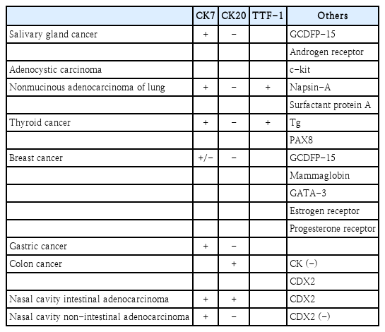
전이 선암종은 면역조직화학염색을 시행하여 선암종의 원발 부위를 유추하는 데 도움을 받을 수 있다. 침샘암의 경우 CK7 양성, CK20 음성을 보이며, 샘낭암종(adenocystic carcinoma)의 경우 c-kit 양성 소견을 보인다[8]. 침샘암은 조직 소견상 유관 유방암(ductal breast cancer)과 비슷한 소견을 보이나, GCDFP-15와 androgen receptor의 존재로 감별에 도움을 받을 수 있다[8]. 폐의 비점액성 선암종(nonmucinous adenocarcinoma)은 조직 소견상 CK7 양성, CK20 음성을 보인다[8]. 그 외에도 폐의 선암종은 TTF-1, napsin-A, surfactant protein A (SP-A)를 포함하여 감별에 도움을 주며, 폐의 선암종과 갑상선암의 감별은 Napsin-A/SP-A (폐의 선암종에서 양성), Tg/PAX8 (갑상선암에서 양성)으로 진단할 수 있다[8]. 갑상선암은 조직학적으로 CK7 양성, CK20 음성과 함께 TTF-1, Tg, PAX8 양성을 나타낸다[8]. 유방암은 주로 CK7 양성, CK20 음성을 나타내나, CK7 음성인 경우도 있다[8]. 유방암은 GCDFP-15, mammaglobin, GATA-3 양성을 보여 감별에 도움이 된다[8]. 하지만 침샘암과 피부암에서도 GCDFP-15 양성을 보이고, 비뇨기계 암에서 GATA-3 양성을 보여 진단에 유의해야 하는데, 유방암에서 estrogen receptor, progesterone receptor 발현을 확인하여 감별할 수 있다[8]. 위암 또한 CK7 양성, CK20 음성 소견을 보이며, 대장암은 CK 음성, CK20, CDX2 양성을 보인다[8,9]. 비부비동의 원발 장형 선암종은 CK7, CK20, CDX2 양성을 보이고, 비장형 선암종은 CK7 양성, CK20, CDX2 음성을 보여 이를 통해 감별에 도움이 된다(Table 1) [10]. 증례 환자에서는 면역조직 화학염색 검사상 CK7 양성 및 CK20 음성, GCDFP-15 양성, TTF-1 음성, CK 양성이 확인되어 침샘암, 유방암, 위암, 비부비동, 비장형 선암종 전이의 가능성을 고려해볼 수 있다.
외비 선암종의 진단과 치료가 보고된 사례가 없었기 때문에 비부비동 선암종의 치료를 참고하였고, 비부비동 선암종은 병변의 수술적 제거 후 종양 절제면의 양성이 확인되거나 고악성도의 종양일 경우 수술 후 방사선 치료 시행을 고려할 수 있다[5]. 본 증례에서는 부비동 전산화단층촬영, 자기공명영상을 촬영하여 주변 구조물로의 침범이 확인되지 않아 우측 코입술주름 하부 조직의 수술적 절제가 가능하였고, 수술 후 항암방사선 치료를 시행하였기 때문에 양호한 예후가 예상되나, 경과 관찰 중에 원발 종양이 발견될 수 있으니 주기적인 추적 관찰과 영상학적 검사가 필요할 것으로 생각된다. 특히 아직까지 선암종이 잘 발생할 수 있는 부위에서 원발암이 발견되지 않았으나, 면역조직화학염색 결과를 고려할 때 침샘암, 유방암, 위암, 비부비동에서 원발 종양이 발견되는지 더욱 주의 깊은 확인이 필요할 것이다. 이를 위해 본 증례 환자에서도 영상학적 검사, 비내시경 검사, 소화기 내시경 검사 등을 정기적으로 시행하고 있다.
Acknowledgements
None
Notes
Author contributions
Conceptualization: Sung Jae Heo. Data curation: all authors. Formal analysis: all authors. Methodology: all authors. Supervision: Sung Jae Heo. Validation: Jae-Hui Kim, Ji Yun Jeong, Sung Jae Heo. Writing—original draft: Boseung Jung. Writing—review & editing: Sung Jae Heo.

