컴퓨터단층촬영에서 비밸브 위치의 정의와 비밸브 협착의 분류
Definition of Nasal Valve Location and Classification of Nasal Valve Stenosis in Computed Tomography
Article information
Trans Abstract
Recently, there has been confusion about the definition of nasal valve stenosis. To resolve this, it is necessary to establish clearly the location of the nasal valve and the definition of nasal valve stenosis. When scrolling the axial view from the top to the bottom, the angle between the upper lateral cartilage and the septum gradually increases, and the vertex becomes rounded. When the lower lateral cartilage is reached, the ceiling of the nasal cavity suddenly becomes higher, so the section immediately before it can be defined as the nasal valve. Nasal valve stenosis is classified as follows. If the difference between the diameters of the narrow and wide nasal valves is less than 1:3, it is normal, whereas if the difference is larger but some space is observed, it is partial stenosis, and if no space is observed, it is complete stenosis.
서 론
비밸브(nasal valve)는 외비밸브(external nasal vavle)와 내비밸브(internal nasal valve)로 분류되지만, 대부분은 내비밸브를 의미한다[1,2]. 비밸브와 비밸브 영역은 다른데, 비밸브 영역(nasal valve area)은 상외측비연골(upper lateral cartilage) 의 하연(caudal margin), 비중격, 하비갑개 전단부, 비강저로 이루어진 단면을 의미하며, 비밸브는 비중격과 상외측비연골의 하연이 이루는 공간을 의미하는데, 서양인은 10-15도 정도의 각을 이루며, 동양인과 흑인은 좀 더 넓다고 알려져 있다[1,2]. 이 비밸브에 협착이 발생하면 심한 코막힘이 발생할 수 있으며, 이를 교정하기 위해서는 비밸브 재건술이 필요하다[3]. 비밸브 재건술은 2014년 9월 19일 신의료기술로 고시되었고, 2016년 4월 18일 비급여 항목으로 지정되어 현재 활발하게 시행되고 있는 것으로 파악된다[3]. 하지만 최근 실손보험회사들이 비밸브 재건술에 대한 치료비 지급을 거부하는 사례가 늘어나고 있는데, 주로 비밸브 협착의 객관적인 입증 여부가 관건이 되고 있다[4].
비밸브 협착의 명확한 진단은 병력 청취, 외비와 비강 내부의 시진, 촉진, 수정 Cottle 검사 등을 종합하여 판단하게 된다[3]. 따라서, 환자를 직접 진찰하지 못한 체 의무기록과 영상 자료만을 근거로 비밸브 협착 여부를 판단하는 것은 현실적으로 매우 한계가 있을 수밖에 없다. 그럼에도 불구하고 비밸브 협착 여부를 판정해야 한다면, CT가 가장 객관적인 자료라고 생각된다. 비록 동적 폐쇄 여부는 알 수 없지만, 자세한 해부학적 구조와 정적 폐쇄 여부는 파악할 수 있기 때문이다. 의무기록의 신체검진 내용은 주관적인 기술이고, 비강 내 내시경 사진은 촬영 위치와 각도에 따라 매우 다르게 보일 수 있으며, 수정 Cottle 검사는 비밸브 협착이 없는 정상인의 경우에도 대부분 양성으로 판정되어 특이도가 떨어진다[5]. 하지만 비밸브 협착의 진단에 있어 공신력 있는 합의나 guideline이 없기 때문에, 심각한 분쟁의 소지가 되고 있다[4]. 여러 논란 중 하나는 CT에서 정확히 어떠한 부위가 비밸브이며, 또 어떠한 경우 협착이라고 정의할 것인가 하는 점이다. 비밸브의 해부학적 정의는 명확하지만, CT에서 구체적으로 비밸브를 확인하기 위한 지표나 방법에 대해서는 의료진들 사이에서도 이견이 있고, 비밸브가 어는 정도 좁아져야 협착이라고 할 것인지에 대한 합의도 없다.
본 연구에서 저자는 비밸브 협착 환자들의 CT를 다각도로 분석하여 정확한 비밸브 위치를 찾고 비밸브 협착을 정의할 수 있는 방법을 제안하고자 하였다. 이는 한 연구자가 독단적으로 결정할 수 있는 사항은 아니지만, 이와 같은 시도가 축적되다 보면 합리적인 합의가 가능할 것으로 생각하였기 때문이다.
방 법
기존에 발표된 비밸브와 인근 구조물의 해부학적 특징들을 근거로 환자들의 CT를 관찰하면서 비밸브를 정확히 찾을 수 있는 방법을 찾고자 노력하였다. 대상 환자들은 본원에서 2023년 6월부터 9월 사이에 비중격 교정술과 하비갑개 절제술을 시행받은 20명(남자 12명, 평균 나이 36.4±7.2세)이었고, 만성 부비동염으로 내시경하 부비동 수술을 동시에 시행받은 경우는 제외하였다. CT를 3차원적으로 분석할 수 있는 프로그램(Ez3D-I, Vatech, Hwaseong, Korea)을 사용하였다. 정량적인 분석이나 통계적 추정은 시행하지 않았다. 본 연구는 후향적 연구로 이미 촬영된 환자의 CT 자료를 분석하였고, 연구대상자를 특정할 수 있거나 그 외 민감정보를 수집하지 않아 본 기관생명윤리위원회의 심의를 면제받았다(KUMC 2023-12-037).
결 과
비밸브를 이루는 상외측비연골의 하연이 해부학적 자세에서 수직면보다는 수평면에 가깝기 때문에 CT의 coronal view 보다는 axial view에서 평가하는 것이 더 적절하다고 생각하여 axial view만을 사용하였다. 비골 부위에서부터 아래쪽으로 탐색하면 상외측비연골이 출현하게 되는데, 비록 CT에서 연골이 관찰되지 않지만, 측면의 비골(Fig. 1A arrows)이 점차 사라지는 것을 보고 추정할 수 있다. 상외측비연골의 상부와 비중격은 매우 급한 예각을 이루며 결합하기 때문에, 두 구조물 사이의 공간도 매우 좁다(Fig. 1A arrowheads). 상외측비연골의 하부로 내려오게 되면 점차 각도가 넓어지지만, 비중격과 이루는 접점은 계속 뾰족하게 유지된다(Fig. 1D-F). 하지만 상외측비연골의 하연 근처에 이르게 되면 비중격과 이루는 접점이 둥글게 변하게 된다(Fig. 1H arrowheads). 이는 기존에 발표된 상외측비연골 해부학 연구 결과와도 일치한다[6]. 비록 접점이 둥글게 변해도 비강 천장의 높이는 거의 일정하게 유지되지만(Fig. 2A and B), 이 부위에서 좀 더 아래쪽을 탐색하면 비강 천장이 갑자기 높아지는데(Fig. 2C and D), 이는 상외측비연골의 위쪽에 위치한 하외측비연골(lower lateral cartilage)로 이행되었기 때문이다. 따라서, 하외측비연골이 출현하기 직전의 상부 CT 단면이 상외측비연골의 하연인 비밸브라고 할 수 있겠다(Fig. 1H and 2B) [1,2,6].

Serial axial view around nasal valve. Fig. A is the uppermost and Fig. L is the lowermost, and the spacing between each cross section is 1 mm. The exact level can be checked in Fig. M. If you explore downward from the nasal bone level, the upper lateral cartilage appears. Although the upper lateral cartilage is not observed on CT, it can be inferred from the gradual disappearance of the nasal bone (Fig. A arrows). The upper lateral cartilage and the nasal septum form a very sharp angle (arrowheads in Fig. A) and the space between the two structures is also very narrow (Fig. A-C). As it descends to the lower part of the upper lateral cartilage, the angle gradually widens, but the apex is still sharp (Fig. D-F). However, when it reaches near the lower border of the upper lateral cartilage, the apex becomes rounded (arrowheads in Fig. H). The blue and orange structures in Fig. M are the virtual upper lateral cartilage and lower lateral cartilage.

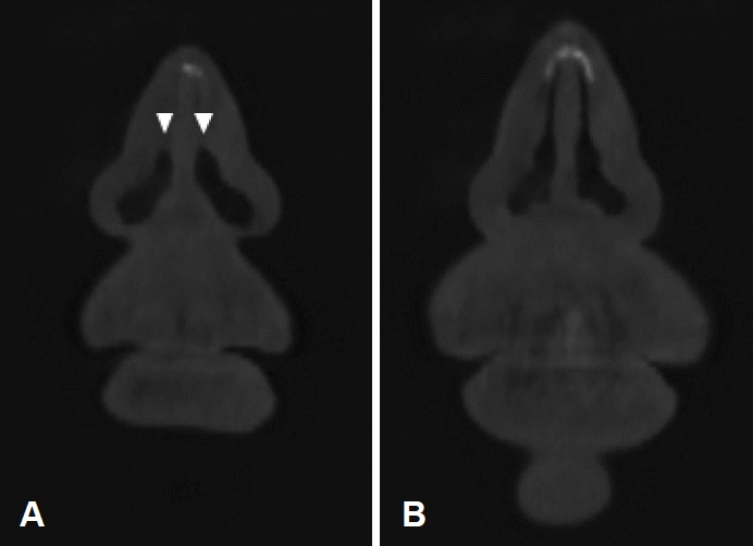
Enlarged axial view around nasal valve. Some photos in Fig. 1 have been enlarged. Fig. A-D are identical to Fig. G-J, respectively. Upon reaching near the lower border of the upper lateral cartilage, the apex becomes rounded but the height of nasal cavity remains constant (Fig. B). Searching more downward, the nasal cavity suddenly becomes elevated (Fig. C and D), as it has been converted to lower lateral cartilage. Therefore, what is observed in Fig. B is the nasal valve. The dotted line represents the horizontal line.
위 설명과 같이 정의한 비밸브 위치에서 분석 프로그램을 사용하여 c oronal v iew를 확인해 보면 Fig. 3A와 같은데, axial view에서 확인되는 둥글게 변한 천장이 마찬가지로 확인된다(arrowheads). 하지만 비밸브 위치를 중심으로 coronal view를 앞뒤로 탐색해 보아도 a xial view에서처럼 비밸브 위치를 명확히 지정할 수 있는 특징이 발견되지는 않았다. Fig. 3B는 비밸브 위치에서 5 mm 후방으로 매우 좁은 공간이 관찰된다.

Nasal valve in coronal view. A: In the coronal plane, the nasal valve (arrowheads) has a rounded nasal vault. B: 5 mm posterior to nasal valve. The nasal cavity is very narrow.
정확한 비밸브 협착을 정의하는 것도 합의가 필요한 사항이다. 과거 상외측비연골과 비중격이 이루는 각도를 측정하 여비밸브 협착을 정의한 연구들이 있었지만, 위에 설명한 것과 같이 비밸브에서 두 구조물이 이루는 각도가 명확히 측정할 수 있는 예각이 아니기 때문에 적용이 어렵다. 결국은 양측의 비밸브의 모양과 넓이를 비교해서 판단하는 방법이 현실적인 분류라고 생각된다[6]. 본 연구자는 Fig. 4A와 같이 양측 비밸브의 모양과 넓이가 거의 동일하면 정상, Fig. 4B와 같이 한쪽 비밸브가 반대쪽에 비해 확연하게 좁지만, 공간이 관찰되면 부분 협착, Fig. 4C와 같이 한쪽 비밸브 공간이 관찰되지 않으면 완전 협착이라는 분류를 제안한다. 부분 협착의 구체적인 정의는 좁은 쪽 비밸브에서 가장 넓은 부분에 수평선을 긋고 좁은 쪽과 넓은 쪽의 비강 폭을 측정하여, 1:3 혹은 더 큰 차이가 나지만 좁은 쪽 비밸브에 공간이 관찰되는 경우이다(Fig. 4D).

Classification of nasal valve stenosis. A: Normal. B: Partial stenosis. C: Complete stenosis. To determine partial stenosis, draw a horizontal line at the widest part of the narrow nasal valve and measure the width of the nasal cavity on the narrow and wide sides (D). If the ratio is greater than 1:3, but some space is observed in the narrow nasal valve, partial stenosis is defined.
고 찰
최근 대한안면성형재건학회에서는 이학적 검사, 내시경 검사, 주관적인 코막힘 평가 등이 비밸브 협착 진단의 필수적인 진단이지만, 추가적으로 CT가 포함될 수 있다고 선언하였다[3]. 또한, CT의 coronal view에서 하비갑개 전단부가 관찰되는 단면 혹은 axial view에서 상외측비연골 하연이 관찰되는 단면이 비밸브라고 간주하고 그 부분에서 비밸브의 단면적을 측정하거나 각도를 측정하는 것을 제안하였다[3]. 하지만, 본 연구자가 3차원 분석이 가능한 CT 프로그램을 이용하여 다수의 환자들을 분석해보면 coronal view에서 하비갑개 전단부의 출현 부위와 비밸브 출현 부위가 반드시 일치하는 것은 아니였으며, 수평면에 가까운 비밸브를 coronal view에서 평가하는 것도 상당히 어려웠다. 또한 a xial view에서 상외측비연골 하연이 관찰되는 단면을 찾는 방법에 대한 구체적인 설명도 없었다. 그 외 CT를 이용한 다양한 연구에서도 비밸브의 정확한 위치를 구체적으로 설명한 경우는 발견하지 못하였다[7-9].
CT에서 상외측비연골과 비중격이 이루는 각도와 공간은 부위에 따라 변화가 크다. 따라서 정확한 비밸브 위치의 합의가 없으면 의사들간에도 비밸브 협착 여부에 대한 판단은 달라질 수밖에 없다. 본 연구자가 제안한 방법을 간략하게 기술해보면 다음과 같다. 즉, CT axial view를 연속적으로 탐색하다 보면, 상외측비연골 상부와 비중격이 이루는 각도가 매우 작고 접점이 뾰족하지만, 상외측비연골의 하연에 가까워지면 점차 각도가 넓어지고 접점이 둥글게 변한다. 여기서 좀 더 아래쪽 단면을 탐색하면 갑자기 비강 천장이 높아지게 되는데, 이는 하외측비연골로 이행하였기 때문이다. 따라서 비강 천장이 높아진 바로 이전 단면이 비밸브가 된다. 이처럼 비밸브는 CT의 특정 단면 하나만을 보고는 판단하기 어렵고 연속된 단면을 반복적으로 탐색해야만 가능하다. 하지만, 비밸브 위치는 CT 단면의 두께, 촬영 각도, 해상도 등에 따라 다소 차이가 날 수 있고, axial view의 평면과 실제 비밸브 단면이 일치하지 않는다는 한계도 있다. 그럼에도 불구하고, 이처럼 구체적인 지침을 적용하면 의료진들 간의 판단 차이를 크게 줄일 수 있을 것이라 생각된다.
Coronal view를 이용해서도 비밸브 위치를 찾아보려고 시도했지만 비밸브를 정의할 수 있는 특징을 발견하지 못했고, 비밸브 위치에서는 비강 천장이 둥글기 때문에 각도 측정도 불가능하였다. 또한 CT의 coronal view를 사용하여 비밸브 협착을 연구한 논문에서 제시한 CT 사진들을 관찰해보면 Fig. 3B와 유사하게 비강 공간이 매우 좁았는데, 이는 실제 비밸브 위치보다 후방이었기 때문이라 추정된다[9].
비밸브의 부분 협착 정의를 좁은 쪽과 넓은 쪽 비강의 폭이 1:3 혹은 그보다 더 큰 차이가 있는 경우라고 정의한 이유는 일반적으로 혈관이나 기도 등 관모양의 구조물에서 직경이 50% 이상 감소하는 경우를 의미있는 협착이라고 정의하기 때문이다[10,11]. 정상 비밸브에서는 양측이 대칭적이기 때문에 비강 폭이 1:1이 되겠지만, 한쪽이 50% 감소하고 반대쪽이 50% 증가하게 되면 비율이 1:3으로 변하게 되며, 차이가 커지면 비율은 더 커지게 될 것이다. 하지만, 이러한 비율이 어떠한 임상적인 중요성을 근거로 제시된 것은 아니며, 단지 판정의 모호함을 줄이기 위해 제안된 것임을 명확히 밝힌다. 현재 실손보험회사들이 비밸브 협착을 완전 협착인 경우만으로 국한시키는 경우가 많은데, 협착(狹窄)의 사전적 의미가 좁아졌다는 뜻이기 때문에 반드시 완전 협착만을 의미한다는 것은 상당히 무리한 해석이며 부분 협착도 포함되는 것이 합당하다고 생각한다. 이를 개선하기 위해서는 비밸브 협착을 정의할 어떠한 기준이 필요하다고 생각되어 제안한 것이다. 명확한 기준은 대한안면성형재건학회에서 논의가 필요한 사항이며, 이후 연구에서 임상적으로 의미있는 협착의 정도가 밝혀진다면 기준에 반영되어야 할 것이다.
본 논문의 모든 내용은 개인적인 제안이며 어떠한 정책이 나 판정의 기준으로 사용될 수 없음을 다시 한번 밝힌다. 다만 비밸브 협착의 판정과 관련되어 매우 혼란스러운 상황이 지속되고 있으므로 이를 해소할 수 있는 구체적 논의가 진행되었으면 하는 바람이 있었고, 마침 “How I Do It”이 개인의 다양한 의견과 방법론을 비교적 자유롭게 표현할 수 있는 공간이기 때문에 이 사이 내용을 제안하게 되었다.
Acknowledgements
None
