부비동 내시경수술 중 발생한 거대 안면 혈종 1예
A Case of a Huge Facial Hematoma During Endoscopic Sinus Surgery
Article information
Trans Abstract
The posterior superior alveolar artery (PSAA) is a branch of the maxillary artery that supplies blood to the lateral wall of the sinus and the overlying membrane. It is located in the lateroposterior wall of the maxillary sinus and is almost intra-osseous, making surgeries there challenging for the risk of injury during routine endoscopic sinus surgery (ESS). Indeed, there is approximately 20% risk of damage in situations such as Le Fort I fracture surgery, maxillary sinus expansion, removal of lesions and inflammation in the maxillary sinus, surgical procedures involving orthognathic surgery and dental implant treatment, as well as in occurrences of fractures and fenestration during surgery, potentially leading to significant bleeding. We present a rare case of facial hematoma due to PASS injury following ESS, with subsequent improvement and no additional complications after treatment.
서 론
부비동 수술은 컴퓨터단층촬영 등 영상의학 기술의 발전으로 부비동의 해부 구조의 확인이 용이해지면서 합병증의 발생을 줄일 수 있었다[1]. 하지만 비강 및 부비동이 좁고 복잡하기 때문에 시야확보의 어려움이 있고 부비동 내시경수술 시 혈관손상을 비롯한 심각한 합병증이 생길 가능성은 항상 존재한다. 비강 및 부비동에는 여러 가지 주요한 혈관 및 작은 갈래의 혈관들이 좁은 공간에 분포하고 있어 이들의 손상으로 다량의 출혈이 생길 수 있고 수술시야를 가려 수술을 힘들게 할 수 있다. 이 중 상악동의 주요 혈관으로는 상악동맥(maxillary artery)에서 분지된 안와하동맥(infraorbital artery), 후상치조동맥(posterior superior alveolar artery, PSAA), 나비입천장동맥(sphenopalatine artery)에서 분지된 측후비동맥(posterior lateral nasal artery)이 있으며, 상악동의 외상이나 수술로 인한 손상으로 심한 출혈을 유발할 수 있다[2].
저자들은 부비동 내시경수술 중 상악동의 동맥 손상이 원인으로 추정되는, 비강내로의 출혈이 없었던 거대 안면 혈종이라는 드문 증례를 경험하였기에 문헌 고찰과 함께 보고하고자 한다.
증 례
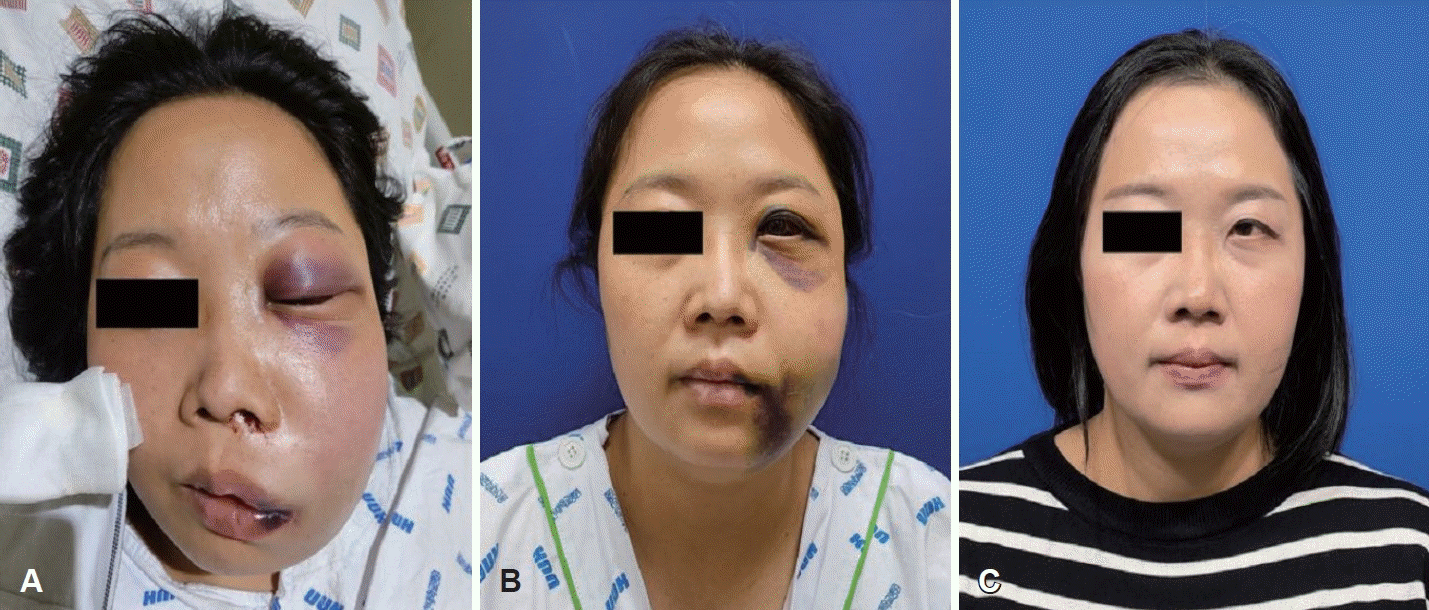
특별한 과거력이 없었던 37세의 여자 환자가 수개월간 지속된 좌측의 만성부비동염으로 타원에서 좌측 부비동 내시경수술을 받은 직후 동측에 통증을 동반한 심한 안면부종이 발생하였음을 인지하였다. 진통제 투여 및 얼음찜질을 시행하였으나 통증과 부종은 점점 더 심해졌다. 수술을 집도한 의사는 저자와의 전화통화에서 수술 중 출혈은 일반적인 부비동 내시경수술 때와 유사하였다고 하였고 얼굴이 갑자기 심하게 붓는 이유를 궁금해하였다. 일단 본원으로 전원을 권유하였고 환자는 1시간 남짓 지난 후 응급실로 내원하였다. 환자는 좌측 얼굴의 심한 부종으로 안면 비대칭 소견을 보였고 좌측 안와 주위의 멍과 안구 돌출이 관찰되기 시작하였다(Fig. 1A). 하지만 좌측 비내시경 검사에서는 수술로 인한 점막의 손상이 있었을 뿐 특별히 심한 출혈양상은 보이지 않았다.

Facial photos of the patient. A: The patient showed severe left facial swelling and ecchymosis with left exophthalmos on the day of the operation. B: The facial swelling had significantly improved by the fourth day of admission. C: The patient had fully recovered without any sequelae three months after surgery.
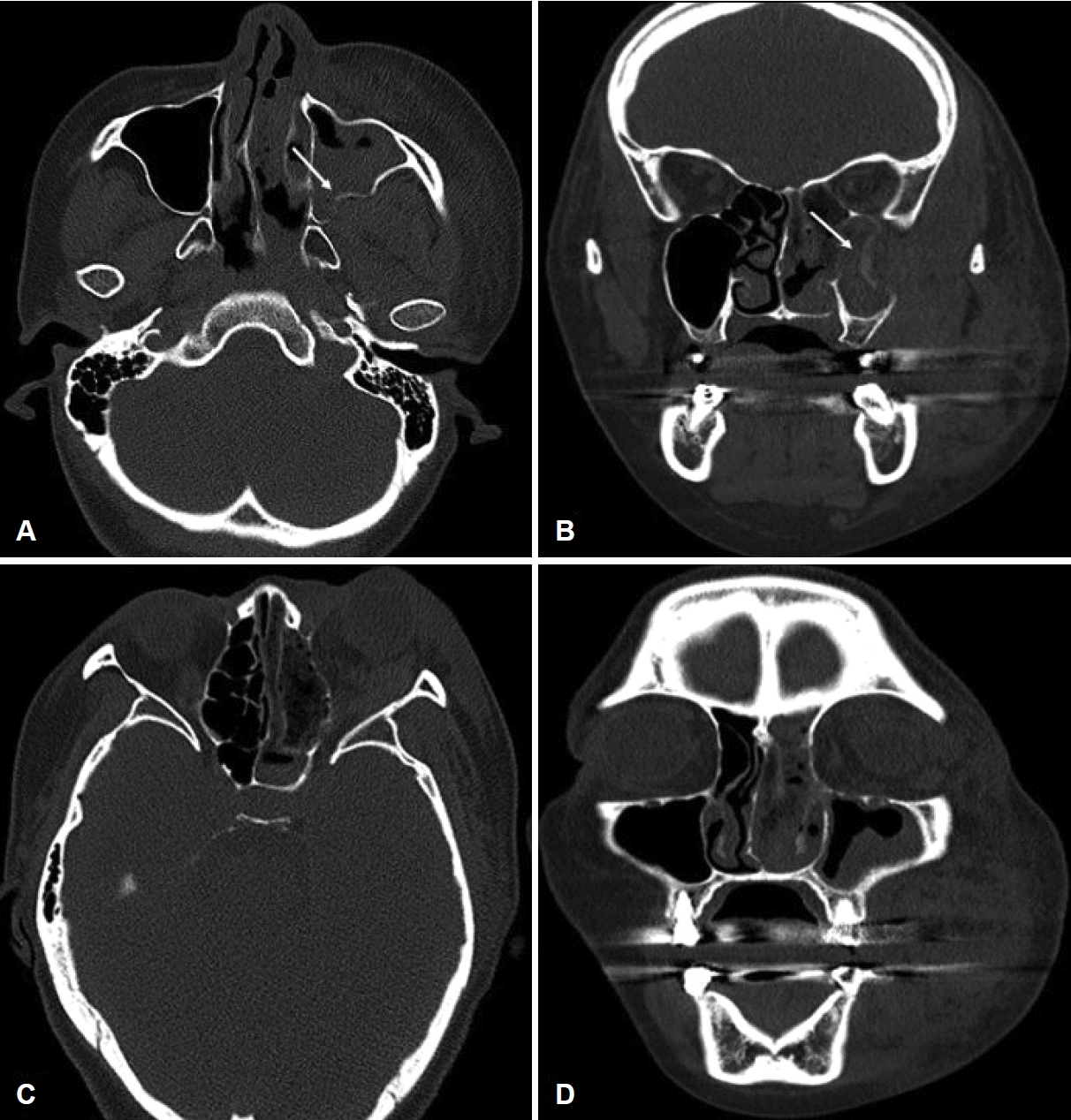
안면 컴퓨터단층촬영검사(CT)상 좌측 상악동 후외측 벽의 골절 및 좌측 안면의 광범위한 연조직 부종과 혈종 소견이 관찰되었다(Fig. 2). 안과에서 시행한 검사상 나안시력은 우측 0.7, 좌측 0.5으로 정상이었다. 안압은 우측 17 mm Hg, 좌측 20.3 mm Hg로 좌안의 안압이 약간 높은 소견을 보였고, 좌측 외안근 운동 검사상 상측 -3, 외측 -2로 운동제한을 보였다. 입원 후의 경과관찰에서 안면이나 경부의 부종이 증가하지 않았고 기도와 혈압, 시력과 안압 등이 정상이어서 혈관 조영술이나 응급수술을 시행하지는 않았다. 혈관손상으로 인한 안면 혈종으로 진단하고 스테로이드(dexamethasone 5 mg qd)와 항생제(ampicillin/sulbactam 3 g qid)를 투여하였으며 안면부종 부위의 얼음찜질을 시행하였다.

CT scans of the patient. A: Axial view of the CT shows a fracture in the posterior wall of the left maxillary sinus (arrow). B: Coronal view of the CT displays a fracture in the lateral wall of the left maxillary sinus (arrow) along with extensive soft tissue edema throughout the entire left face. Axial (C) and coronal (D) views of the CT show intact bone state around of the orbital wall.
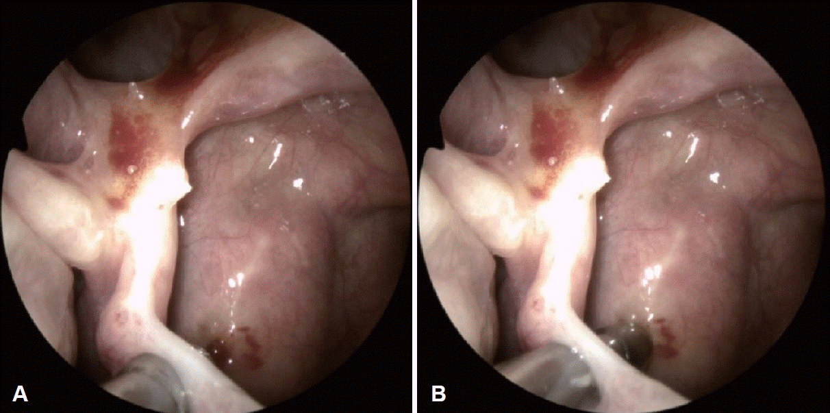
입원 2일째 안과검사에서 안압은 우측 14.7 mm Hg, 좌측 14.4 mm Hg로 감소되었으며, 외안근 운동 검사에서 상측 -0.5, 외측 -0.5로 운동제한을 보였으나 내원당일보다는 안구움직임이 호전된 것을 확인할 수 있었다. 이후 안면 부종 및 혈종크기가 전반적으로 감소하여(Fig. 1B) 입원 4일째에 dexamethasone 투여를 중단하였으며 추가적인 합병증이나 출혈이 없어서 입원 6일째 퇴원하였다. 퇴원 후 1개월째 외래 경과관찰 시 시행한 비내시경 검사상 좌측 상악동 개구부 아래로 점막의 천공과 상악동 후벽 점막의 멍이 관찰되었다(Fig. 3). 퇴원 후 3개월째 경과관찰에서도 합병증 없이 정상으로 회복되어 치료를 종결하였다(Fig. 1C).
고 찰
1985년 Kennedy, Stammberger 등에 의해 부비동 내시경 수술이 처음 알려진 후 부비동 수술은 대부분 비내시경을 사용해 이루어지고 있다[3]. 또 1990년 초부터 악관절 수술 목적으로 개발된 정상 조직의 손상을 최소화할 수 있는 회전식 흡입기(microdebrider)가 부비동 수술에 이용되게 되면서 수술 중 출혈에 의한 시야 장애와 정상 비강점막조직의 손상을 최소화할 수 있게 되었다[1,4]. 컴퓨터단층촬영 등 영상의학 기술의 발전 역시 병변의 범위와 주변부의 해부 구조의 변화에 대해 분명한 정보를 제공하여 수술에 대한 안정성을 증가시켰고 합병증 발생비율을 감소시켰다[5]. 그럼에도 불구하고 부비동 수술에 따른 합병증은 여전히 발생하고 있는데 그 비율은 0.3%-22.4%까지 보고되고 있고, 수술 중에 발생하는 심한 출혈의 경우에는 0.36%-3.1% 정도로 보고되었다[6-8].
상악동 주위에 분포하여 수술 중 출혈을 초래하는 대표적인 혈관은 측후비동맥(posterior lateral nasal artery)으로 나비입천장동맥(sphenopalatine artery)에서 분지되어 비강의 외측벽과 중, 하비갑개에 혈액을 공급한다. 상악동 자연공개창술(middle meatal antrostomy) 시행 시 개창술을 후방으로 확장할 때 손상 받기 쉬워 잦은 출혈이 일어난다[9]. 한편 상악동의 상벽에 위치하는 안와하동맥(infraorbital artery)의 경우에는 상악동의 상벽, 즉 안와하벽이 내측에서 외측으로 경사지고 벽이 얇아 외상이나 수술 중 상벽의 조작으로 손상을 받아 출혈이 생길 수 있다[10]. 이에 반해 후상치조동맥(PSAA)은 상악동의 측하방의 골내(intra-osseous)에 대부분 위치하고 있어 통상적인 비내시경을 통한 부비동 수술 시 접근이 어려울 뿐만 아니라 흔하게 손상을 받는 위치가 아니기에 출혈이 잘 발생하지 않는다. 일반적으로 Le Fort I 골절 수술, 양악 수술 및 치과 임플란트 치료 등과 같은 상황에서 약 20%의 손상가능성이 있고 이로 인해 심한 출혈을 유발할 수 있다[2,11].
하지만 이번 증례의 환자의 경우에는 후상치조동맥의 손상이 거대 혈종의 원인으로 추정된다. 상악동의 상벽인 안와 하벽에 위치한 안와하동맥(infraorbital artery)의 손상에 의한 출혈도 생각해볼 수 있으나 만약 이곳에서 출혈이 발생하였다면 혈종이 상악동 뒤쪽인 익구개와 혹은 안와하부에 발생했을 것이다. 하지만 혈종이 안면 외측으로 크게 발생하였고, CT에서 발견되는 좌측 상악동 후외측의 골관 하단에서부터 뼈 능선(bone crest)에 이르는 골절소견은 그 부위를 지나가는 후상치조동맥의 손상을 추정하게 한다. Takahashi 등[12]은 상악 제3대구치에 국소마취를 하던 과정에서 발생한 후상치조동맥의 손상에 대한 증례를 보고하였는데 혈종으로 인해 심한 안면 부종이 발생한 환자의 사진이 본 증례의 사진과 유사하였다. 그 증례의 경우 경부의 부종과 통증이 악화되어 혈관조영술 및 동맥색전술을 시행하였다.
점막의 부종과 가피가 사라진 수술 후 한달 째의 외래 경과 관찰에서 상악동 자연공 아래로 점막의 천공이 관찰되었는데 구부러진 석션팁(curved suction tip)을 넣었을 때 끝이 닿는 부위와 골절 부위에 생긴 멍자국이 일치하였다(Fig. 3). 이를 통해 술자가 curved suction이나 curved shaver tip을 이용해 무리하게 상악동의 천문(fontanelle)에 구멍을 뚫으려고 하다가 tip이 갑자기 깊이 들어가면서 상악동 후벽에 골절을 일으킨 것으로 추정한다. 골절로 인해 상악동 후벽에 위치한 동맥이 손상되었지만 골절편이 체크밸브 역할을 하여 상악동으로의 출혈은 발생하지 않았고 대신 얼굴의 거대 혈종을 만든 것으로 추측한다.
이번 증례의 경우에는 다행히 지속적인 출혈을 의심할만한 얼굴 부종의 증가나 혈압저하 등이 없어서 추가적인 치료는 필요하지 않았다. 하지만 얼굴이나 경부의 부종이 악화되는 소견을 보인다면 혈관조영술과 동맥색전술이 필요하다. 또한 안압상승으로 인한 시력저하가 초래되는 경우에는 안와감압술이 동반되어야 한다.
후상치조동맥은 익상구개와(pterygopalatine fossa) 내의 상악동맥(maxillary artery)에서 분지되는 혈관이다[13]. 하측 두엽표면(infratemporal surface)에서 하방으로 내려가며, 상악의 후측방에 있는 구멍을 통해 안쪽으로 들어가 골관을 통해 상악동 측하방을 지나간다[14]. 후상치조동맥은 99.4%에서 존재하는데 골내(intra-osseous)에 존재하는 경우가 84.2%, 점막하가 12%, 외측 부비동 벽의 외부 피질(outer cortex of lateral sinus wall)이 3.2%를 각각 차지하는 것으로 보고되었다[15]. 또 Park 등[16]이 시행한 혈관 위치에 관한 연구에서 골관 하단에서부터 뼈 능선(bone crest)까지의 거리가 치악군에서는 소구치에서 20.62±3.05 mm, 대구치에서 17.50±2.84 mm로 측정되었고, 무치악군에서는 소구치에서 18.83±2.79 mm, 대구치에서 15.50±1.64 mm로 측정되었다.
부비동 내시경수술 시 혈관손상으로 인한 출혈과 이로 인한 합병증의 가능성이 늘 존재하므로 술자는 주요 구조물의 해부와 더불어 혈관의 주행을 숙지해야 한다. 따라서 술전 영상검사를 통한 확인이 필수적이고, 혈관 주변의 병변을 제거할 때는 직접 눈으로 확인하며 조심스럽게 기구를 조작하며 제거해야 한다. 만약 시야 확보가 어렵다면 상악동 입구를 좀 더 확장하거나 견치와 천공(canine fossa puncture) 및 전누관접근(prelacrimal approach) 등과 같은 술식을 통해 넓은 수술시야 및 기구사용을 위한 공간을 확보할 수 있고 이를 통해 수술 중 발생할 수 있는 합병증을 줄일 수 있을 것으로 생각한다.
결론적으로, 비강 및 부비동 내에는 여러 가닥의 혈관이 주행하고 있으며 이 중 동맥손상에 의한 출혈은 출혈량이 많아 심각한 결과를 초래할 수 있다. 저자들은 부비동 수술로 인해 생긴, 출혈을 동반하지 않은 심한 안면 혈종이라는 드문 증례를 보고하면서 비부비동의 해부학 구조를 잘 파악하고 무리한 기구 조작을 피하며 방심하지 않고 항상 긴장하는 자세의 중요성을 다시 한번 강조하고자 한다.
Acknowledgements
None
Notes
Author contributions
Conceptualization: Tae-Hoon Lee. Data curation: Jae Won Jang. Investigation: Jae Hyun Kim. Project administration: Tae-Hoon Lee. Resources: Sang Hyok Suk. Supervision: Tae-Hoon Lee. Visualization: Jae Won Jang. Writing—original draft: Jae Hyun Kim. Writing—review & editing: Tae-Hoon Lee.

