Assessment of Nasal Dimensions at the Pyriform Aperture and Mid-Nose in Neonates and Infants With Nasal Obstruction
Article information

Abstract
Background and Objectives
Neonatal nasal obstruction can lead to significant morbidity. We carried out this study to investigate the radiological measurements of nasal dimensions that are correlated with the diagnosis of congenital bony stenosis, and to present a management strategy for neonatal nasal airway obstruction.
Subjects and Method
A total of 54 patients were included for the study. We conducted CT scans for all the patients to assess the cause of nasal obstruction. Among these patients, 27 individuals had nasal obstruction caused by stenosis at the level of pyriform aperture (PA) or mid-nose (MN), while the remaining 27 experienced nasal obstruction due to different causes. Radiological measurements were conducted and compared between these two groups. Operative data and follow-up information obtained for a period of 6 months were reviewed.
Results
Patients requiring surgery for the treatment of nasal stenosis exhibited a PA with the average width of 6.2 mm ([SD±1.7]; median [IQR] 6 [5-6.5]), and a MN with the average size of 5.1 mm ([SD±0.9]; median [IQR] 5 [4.4-6]). Infants experiencing nasal obstruction due to other reasons had an average PA measuring at 11.7 mm ([SD±1.9]; median [IQR] 12 [11-13]) and MN at 8.1 ([SD±1.3]; median [IQR] 8.2 [7-9]).
Conclusion
Clinical, endoscopic, and radiological data together can help determine whether patients with nasal stenosis need surgery. We found that PA stenosis is often associated with MN level stenosis.
Introduction
Nasal obstruction in a neonate is a life-threatening condition as it can cause significant respiratory distress. It is crucial to promptly diagnose and treat this condition to ensure the well-being of the infant. Causes include mucosal inflammation and bony narrowing. Choanal atresia is the commonest cause of complete bony obstruction. Congenital bony nasal stenosis at the level of the pyriform aperture (PA) and/or mid-nose (MN) cavity without atresia, although rare, can cause significant symptoms such as cyanosis, feeding difficulties, failure to thrive and obstructive sleep apnea [1].
Clinical and radiological description of pyriform aperture stenosis (PAS) has been the focus of many previous studies [2-4]. Our understanding of the pathology and impact of this abnormality has evolved during the last two decades [5-8]. However, certain aspects are still yet to be fully studied such as indications for surgery, the best surgical technique, factors affecting outcome and causes of failure and/or restenosis.
This study aims to provide a comprehensive evaluation of neonates and infants with symptoms of nasal obstruction, focusing on the role of radiological assessment in identifying those who may require surgery. Additionally, we present our experience as a tertiary pediatric hospital in addressing this issue, offering a practical approach for managing these cases effectively.
Subjects and Methods
This study focused on evaluating children with respiratory distress caused by nasal obstruction. The inclusion criteria encompassed all patients admitted for assessment of significant nasal obstruction between June 2020 and June 2023. The significance of nasal obstruction was determined by several factors, including the need for intubation or non-invasive ventilation, failure to thrive, recurrent cyanosis, obstructive sleep apnea, and the response to conservative measures such as nasal saline, steroids, and frequent suction. Total number of patients was 82. We then excluded children over 6 months (n=15), recurrent cases (n=4), and those with concurrent airway anomalies (n=9). ENT examination was conducted, including clinical examination as well as flexible nasal endoscopy using both 4 mm and 3.4 mm scopes. Flexible endoscopy was done by the author and the 3.4 mm scope was tolerated by all neonates with an assistant providing head stabilization during the procedure. In addition, CT scans of the nose and paranasal sinuses were performed. The study population was divided into two groups based on specific criteria: group A and group B. Detailed analysis involved reviewing CT images along with preoperative assessment, operative notes, and follow-up data.
Group A
Group A consisted of children who experienced respiratory distress due to nasal obstruction caused by congenital bony stenosis without atresia or mucosal pathology. These cases underwent surgical correction of the bony stenosis either at the level of PA or MN or both. Table 1 provides information on patients’ demographics.

Patient demographics, CT dimensions, and the procedure patients had in group A to address nasal stenosis
Group B
Group B consisted of patients who had respiratory distress due to nasal obstruction and either showed improvement with conservative treatments or underwent surgery for issues unrelated to congenital bony stenosis. A comparison was made between group A and group B based on radiological measurements of the nasal cavity at two levels: PA and MN.
Radiographic measurements
CT measurements were conducted in the axial view for each patient. Axial measurements were obtained at the PA and MN regions. The landmarks used for measurement included the bony lateral nasal wall (nasal process of maxilla) to the contralateral bony lateral nasal wall at the level of the PA (Fig. 1). Additionally, a measurement was taken at midnosal area, which represents the narrowest middle mucosal width between both inferior turbinates (ITs) (Fig. 1). Table 1 displays nasal cavity dimensions within group A, while Table 2 illustrates nasal cavity dimensions within group B.
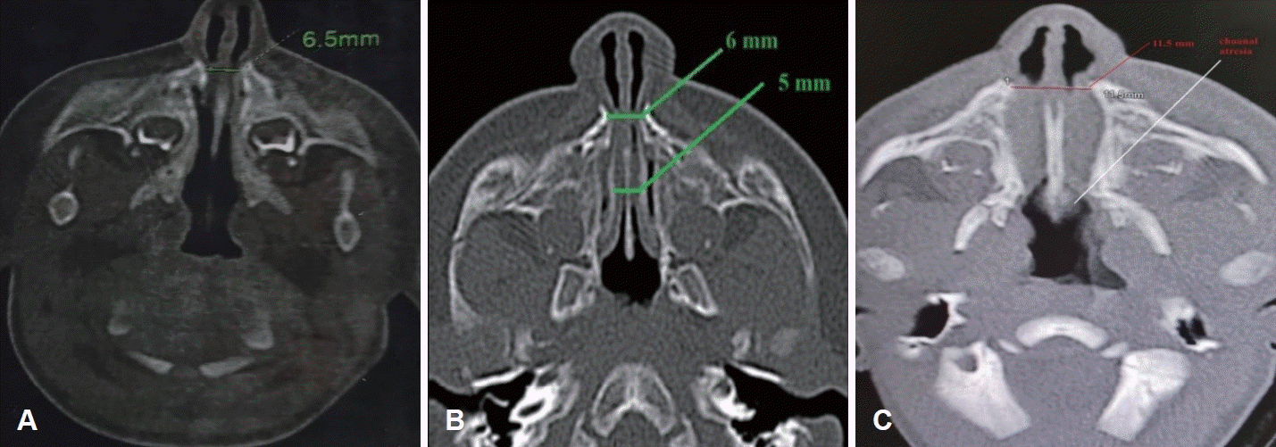
CT assessment of nasal dimensions. A: CT scan of a patient in group A with pyriform aperture (PA) width of 6.5 mm. B: CT scan of a patient in group A with PA and mid-nose of 6 mm and 5 mm respectively. C: CT scan of a patient in group B with PA width of 11.5 mm and evidence of choanal atresia.

Group B patients who had CT for evaluation of nasal obstruction
Surgical intervention
The criteria used to proceed with surgical treatment to correct congenital nasal stenosis (group A) encompassed the combination of severe airway obstruction symptoms, endoscopic confirmation of narrow nasal passages, and radiological measurements indicating narrow nasal dimensions without complete nasal atresia. Surgical intervention included one or a combination of three procedures: (A) a sublabial approach and drill out of the side wall and floor of the pyriform fossa on both sides. (B) Gentle nasal dilatation with Hegar dilators and IT out fracture or trimming. (C) Nasal stents (endotracheal tube) which were left in place for 1-6 weeks. All surgical procedures were done by one surgeon: the author. Table 1 shows summary of surgical interventions done for patients in group A. Regarding group B, thirteen patients had choanal atresia repair, eight had adhesion release and six received medical treatment for neonatal rhinitis.
Data analysis
We conducted a comparison of the dimensions of bony nasal structures in patients with symptomatic nasal stenosis requiring intervention or who did not respond to conservative measures (group A) and those undergoing radiological evaluation for nasal obstruction unrelated to congenital bony nasal stenosis (group B). We thoroughly examined the medical records, including operative notes, interventions, 6-month follow-up data, and any complications.
Statistical methods
Data was entered and statistically analysed on the Statistical Package of Social Science Software program, version 25 (IBM SPSS Statistics for Windows, Version 25.0; IBM Corp., Armonk, NY, USA). Data for the quantitative variables was presented using range, mean±standard deviation (SD) and median with the interquartile range (IQR). The 95% confidence interval was reported for the median value. Comparison between groups for was performed using Mann-Whitney test (due to lack of normality of data). Receiver operating characteristics (ROC) curve analysis was performed to explore the discriminant ability of both PA and MN measures in differentiating group A cases using the most appropriate cut-off point by Youden Index. Inter-rater reliability analysis between PA and MN measures for each group separately was conducted using the intra-class correlation coefficient by two-way mixed modelling of consistency sub-type. p-values less than or equal to 0.05 were considered statistically significant [9].
Ethical considerations
This study is a retrospective review of patients treated at Cairo University Children Hospital. The study was approved by the scientific committee of the department and by the research ethics committee of faculty of medicine, Cairo University (N-295-2023). All data are anonymous and it’s impossible to identify patients from this report. I confirm that the procedures followed were in accordance with the ethical standards of the responsible committee on human experimentation and with the Helsinki Declaration of 1975, as revised in 1983.
Results
Group A
Group A consisted of a total of 27 patients, with a distribution of 11 males and 16 females. The age range in this group varied from 1 to 148 days, with an average age of approximately 48 days. Co-morbidities observed among these patients included solitary median maxillary central incisor syndrome, dextrocardia with situs inversus, cardiomyopathy, craniosynostosis, pulmonary hypertension, imperforate anus, Apert syndrome and micrognathia.
Group B
There were 27 patients in this group: 14 females and 13 males. Age range 2-90 days with an average of 30 days. The main nasal issues observed in the patients included bilateral choanal atresia in 13 cases, nasal adhesions in 8 cases, and rhinitis in 6 cases.
Radiological measurements

Statistical results and significance
Table 3 provides a comprehensive comparison of both groups and demonstrates a statistically significant difference in PA and MN measurements between group A and B, indicated by a p-value of less than 0.001 [9]. The data presented in Fig. 2 (box plot) illustrates the distribution of measures for PA and MN width between group A and group B. In addition, Fig. 3 provides an ROC curve analysis to assess the discriminant ability of PA and MN measures in differentiating between the two groups. The results displayed in Table 4 indicate that both measures have an area under the curve greater than 0.9, indicating excellent discrimination between group A and group B. The reliability assessment (inter-rater agreement) of PA and MN measures in each group separately demonstrated that intraclass correlation coefficient values for reliability between PA and MN measures for each group separately are between 0.5 and 0.75 indicating a moderate degree of reliability (Supplementary Table 1).
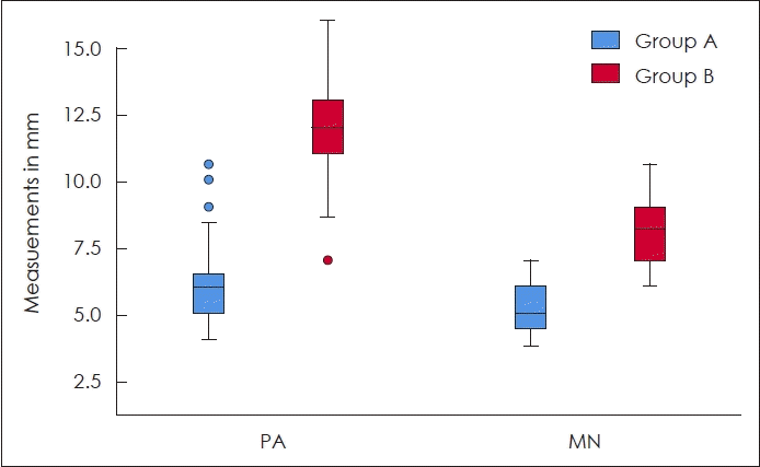
Box plot showing the distribution of pyriform aperture (PA) and mid-nose (MN) measures between both groups.
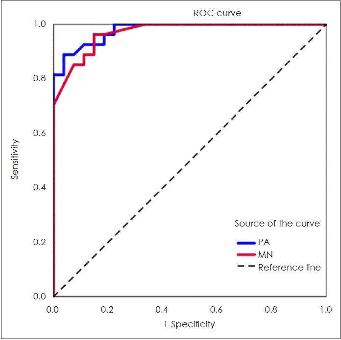
Receiver operating characteristics (ROC) curve analysis to explore the discriminant ability of pyriform aperture (PA) and midnose (MN) measures in differentiating group A from group B.

Area under the curve for pyriform aperture and mid-nose measurement
Operative findings
Surgical intervention for nasal stenosis was tailored based on the dimensions revealed by CT scans and intraoperative findings. Table 3 provides an overview of the various surgical procedures conducted for group A patients. For cases with CT evidence of PAS, a sublabial approach was used to perform drilling of the PA under endoscopic control (Fig. 4). In addition to addressing the PA, it was also observed that most cases required dilatation of the MN. On the other hand, there were instances where isolated MN stenosis was present in certain cases (number 5, 8, 11, 14, 21, and 23), necessitating surgical widening of the MN without addressing the PA. Temporary stenting for a period ranging from 1-6 weeks was performed for most cases.
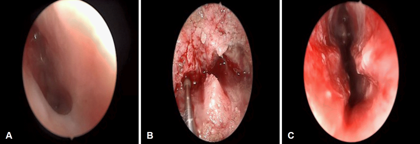
Endoscopic nasal intraoperative photos of a case with PAS. A: Left nasal cavity with severe PAS and adhesions and the anterior end of inferior turbinate is not fully seen. B: Intraoperative photo where the lateral wall of the PA is drilled. C: Post operative view with significant improvement of the cross-sectional area of the nasal passage.
Complications
All cases in group A showed significant improvement in their airway obstruction and nasal symptoms. However, further intervention was required for cases 2, 3, 5, 6, and 10 due to either symptomatic restenosis or complications related to stenting such as nasal adhesion. Two cases (6 and 10) experienced septal perforation after the removal of the stent and exhibited evidence of columellar width retraction.
Discussion
A patent nasal passage is essential for the normal breathing of neonates. The paediatric ENT surgeon needs to establish the degree and level of obstruction and determine the most appropriate intervention. Fig. 5 shows the suggested flowchart for the management of neonates and infants with nasal obstruction.
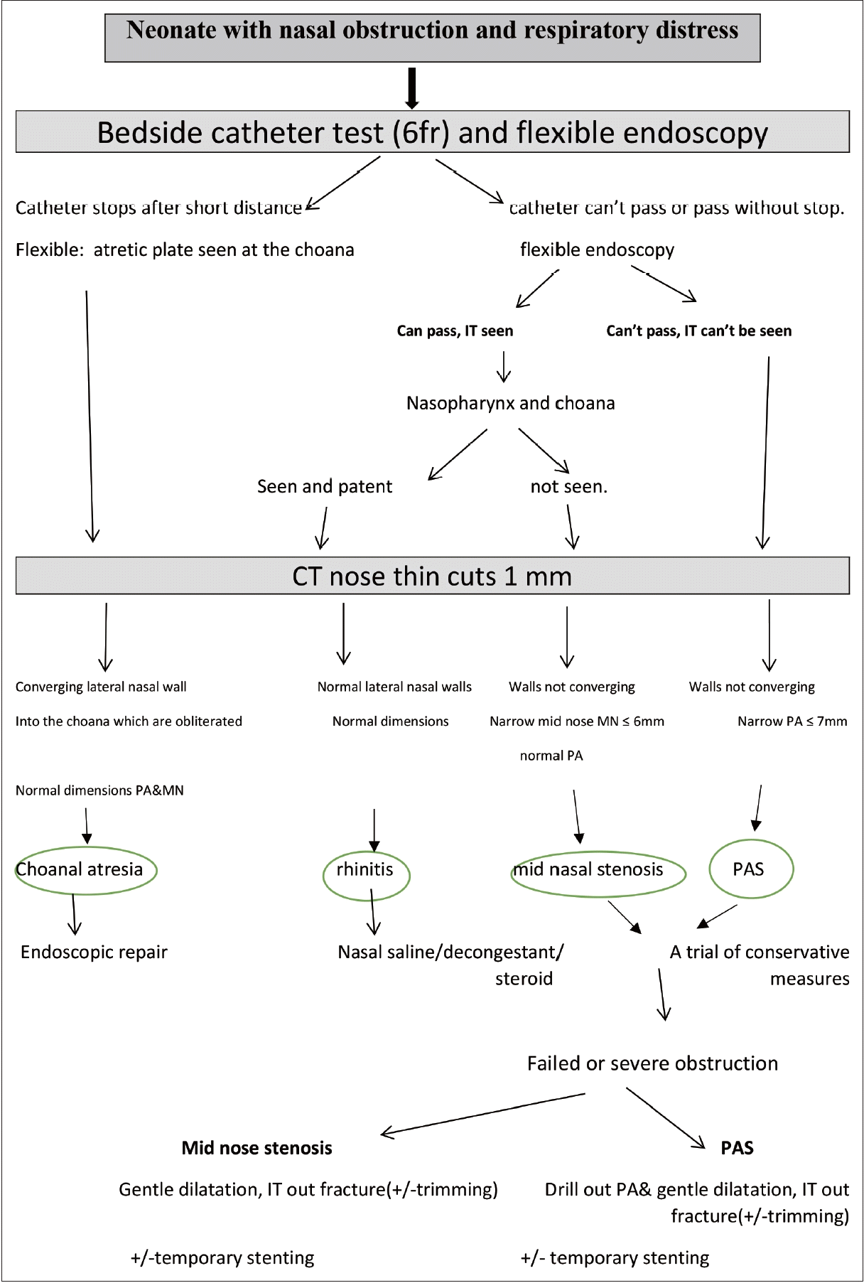
Flowchart for the management of neonatal nasal obstruction. Note that CT scan can be omitted if neonatal rhinitis is suspected particularly if there is improvement with nasal steroid drops. IT, inferior turbinate; PA, pyriform aperture; MN, mid-nose; PAS, pyriform aperture stenosis.
Surgical correction of nasal stenosis poses more challenges when there is a global narrowing rather than a single site, according to Graham, et al. [10] Cases associated with Pierre Robin sequence are best treated with a nasopharyngeal airway serving both as a reliable airway bypassing glossoptosis-induced obstruction and acting as a temporary stent after surgical intervention.
In terms of cut-off value for surgical treatment based on PA width, Visvanathan and Wynne [11] highlighted that a PA width of <5 mm almost always requires surgical correction although this wasn’t statistically proved in their study including 10 patients. Wormald, et al. [12] mentioned that a PA width less than 5.7 mm is 88% sensitive and specific in predicting the need for surgery. On the other hand, Gonik, et al. [13] concluded that there is no correlation between PA size and the requirement for surgery. Interestingly, they found that patients who underwent surgery had an average PA size of 5.71 mm compared to conservative management cases with an average of only 4.83 mm. In our analysis, it is possible that the variability in a child’s ability to cope with nasal stenosis and inconsistencies in measurement techniques may account for this finding. In this study, patients with severe symptoms who required surgery had an average PA width of 6.2 mm (SD=1.7) and a MN width of 5.1 mm (SD=0.9).
In a study of 62 neonates and infants [14] who did not have nasal obstruction, the mean and range of PA and MN were 13.24 (8.8-17.2) and 7.9 (4.9-13.5) respectively. In an article by Pollaers, et al. [15] who used one hundred controls, the mean PA width for cases was 5.6 mm (range 4-7.5 mm) compared to a mean width of 12.8 mm (range 9.4-16.6 mm) for normal controls. The authors found that the bony nasal cavity was statistically significantly narrower in cases compared to controls along 75% of the distance of the nasal cavity. Those findings are consistent with our results.
According to Shah, et al. [16], maternal diabetes mellitus has been identified as a potential factor that may increase the risk of PAS. In our study, three patients who had this same maternal comorbidity required mechanical ventilation for respiratory support.
Mid-nasal stenosis is a lesser-known cause of nasal obstruction in newborns. The author observed that despite widening the PA by drilling the lateral wall, there was still posterior stenosis due to the anterior end of the IT remaining more medial than the drilled area. This finding was confirmed by preoperative CT measurements of both the PA and MN.
Our findings indicate that PAS is not always limited to a single-level abnormality. The narrowing or thickening of the pyriform processes, formed by the nasal processes of the maxilla, and connecting with the IT at the conchal crest, can cause a medialization of the IT and the lateral nasal walls. Hence, we believe that cases with PAS have a degree of mid-nasal stenosis as well.
These findings were also noted in a small study [17] of four patients with severe PAS who underwent nasal dilatation without bony drill out. However, two patients required additional dilatations at short intervals. Devambez, et al. [18] stated that partial inferior turbinectomy was necessary in seven out of nineteen cases with PAS, which aligns with another study’s findings [19]. Reeves, et al. [6] compared seven cases of PAS with thirteen controls in infants less than a month and stated that the anterior three-fourths of the nasal cavity is stenosed in cases of PAS. Equivalent results were reported by another study [15].
In this series, the author observed that in the presence of PAS, medialized IT accounts for mid-nasal stenosis. Nevertheless, isolated mid-nasal stenosis with the normal endoscopic and radiological evaluation of PA, in the context of a neonate with nasal obstruction, is probably due to hypertrophy of the IT rather than a medialized site of insertion. In this series, six patients (5, 8, 11, 14, 21, and 23) were identified with mid-nasal stenosis and normal PA width.
Raghavan, et al. [20] in 2003 described a case of congenital mid-nasal stenosis where he found a tight stenosis on both sides about 3 cm from the PA. This was dilated with Hegar dilators and a stent was inserted and left for 8 weeks. He concluded that mid-nasal stenosis should be suspected in children with nasal obstruction, who have failed medical management and without PAS.
According to a study by Syed, et al. [21], bedside fibreoptic evaluation can be used to assess neonates based on the appearance of the middle and ITs. This evaluation method is applicable in diagnosing PAS, where severe cases may not allow visualization of the anterior end of the IT.
A considerable number of patients with nasal blockage caused by stenosis in the mid-nasal region was documented by Patel and Carr [22]. In their study, all cases received medical management, but it took an average of 126 days for symptoms to resolve and only one-third underwent imaging. The report did not mention any measurements obtained from CT scans. Some research studies [22,23] have identified unilateral mid-nasal stenosis as a potential cause of nasal obstruction in newborns. However, it is unclear why infants with unilateral stenosis experienced such severe symptoms.
Through our observations, we have identified three subtypes of MN stenosis: 1) an isolated abnormality primarily caused by congenital hypertrophy of the ITs; 2) MN stenosis associated with PAS resulting from a narrow distance between the lateral nasal walls; and 3) As a part of a craniofacial abnormality such as mid facial hypoplasia due to abnormalities in the cranial bones leading to global stenosis.
In conclusion, careful radiological assessment is recommended for diagnosing congenital nasal stenosis. The average PA width is 6.2 mm, and the MN width is 5.1 mm in severe cases requiring surgery. Mid-nasal stenosis is often linked to PAS, and it is advisable to address both levels during surgical treatment. Our findings suggest that mid-nasal stenosis can be an isolated anomaly, associated with PAS or a part of global nasal stenosis.
Supplementary materials
The Supplement is available with this article at https://doi.org/10.3342/kjorl-hns.2024.00290.
Acknowledgements
None
Notes
Availability of Data and Materials
The datasets during and/or analysed during the current study available from the corresponding author on a reasonable request.