경외이도 내시경 등골 수술: 서술적 고찰
Transcanal Endoscopic Stapes Surgery: A Narrative Review
Article information
Trans Abstract
Stapes surgery has evolved from stapes mobilization to stapedotomy, which was initially performed using a surgical loop and is now done using an operating microscope. The improvement of endoscopic systems is attributed to the advancement of optical and medical engineering technologies, and has made endoscopic ear surgery a popular choice for various middle ear surgeries. In particular, endoscopic stapes surgery (ESS) has an advantage over the conventional microscopic stapes surgery (MSS) because it provides a wide-angled and magnified view of the middle ear structures, including the stapes, during surgery. All stapes surgical procedures can be performed exclusively using the transcanal endoscopic approach. Moreover, the postoperative hearing outcomes of ESS are comparable to those of conventional MSS. However, being a one-handed procedure with a steep learning curve, ESS can be an alternative to the conventional MSS, provided that the surgeon understands the advantages and disadvantages and is properly trained.
Introduction
Transcanal endoscopic stapes surgery (ESS) was introduced approximately a quarter of a century ago. In 1999, Tarabichi [1] first reported the results of ESS in patients with otosclerosis. The author performed 13 cases of stapedectomy using an endoscope. There were no postoperative complications, such as sensorineural hearing loss, and air-bone gap closure within 10 dB was achieved in six of seven patients with an available follow-up 1 year after surgery [1]. In 2000, Poe [2] performed stapedioplasty using a laser endoscope on eight patients with otosclerosis. The fixed anterior third of the stapes footplate was cut with a laser and removed without inserting a stapes prosthesis. The eight patients showed an average air-bone-gap improvement of 14.8 dB with no postoperative hearing loss [2]. Despite the successful results of ESS, it has not gained popularity because of its difficulty. In recent years, however, ESS has become increasingly popular as our understanding and experience have accumulated in various middle ear surgeries such as tympanoplasty, ossiculoplasty, and middle ear tendon lysis [3-6]. This review describes the surgical procedures, advantages, disadvantages, and outcomes of ESS.
Surgical Procedures
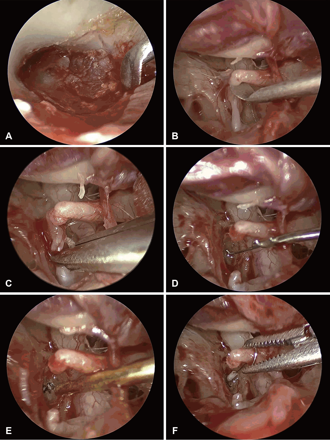
The procedure begins with a four-quadrant ear canal injection of 1:100000 epinephrine, with care taken to avoid bullae formation. Two vertical incisions are made for the tympanomeatal flap: the first is anterior to the lateral process of the malleus (at the 2 o’clock position for the right ear and 10 o’clock for the left), and the second is at the 6 o’clock position. This wide incision design allows for the assessment of concomitant malleus fixation. A horizontal incision connects the two vertical cuts approximately 8-10 mm lateral to the tympanic membrane, which ensures adequate exposure for the subsequent removal of the posterosuperior bony canal wall (Fig. 1A). To elevate the flap while minimizing the risk of injury to the chorda tympani nerve, entry into the middle ear cavity begins inferiorly (at the 6 o’clock position) and proceeds posterosuperiorly. Once the flap is elevated, the chorda tympani nerve is identified. The posterosuperior bony canal wall is then removed to fully expose the incudostapedial joint, stapes, and oval window niche, taking care to preserve the nerve. The mobility of the ossicular chain is assessed. After confirming stapes fixation, the incudostapedial joint is separated (Fig. 1B), and the lateral ossicular chain is reassessed for any concomitant fixation. The stapedius tendon is then cut. The stapes suprastructure is subsequently removed by first dividing the posterior crus with crurotomy scissors (Fig. 1C) or a laser, followed by fracturing the anterior crus inferiorly toward the promontory with a right-angled pick. This downward motion is a critical step to prevent inadvertent mobilization or subluxation of the footplate. The functional length between the stapes footplate and the medial border of the long process of the incus is measured using a measuring rod (Fig. 1D). A fenestra is created on the stapes footplate using a laser and/or a microperforator (Fig. 1E). The piston prosthesis, sized according to the measured functional length, is placed between the fenestra and the long process of the incus (Fig. 1F). If the long process of the incus is eroded, a malleostapedotomy is performed. Finally, the fenestra is sealed with soft tissue or fibrin glue, and the tympanomeatal flap is repositioned.

Surgical steps of endoscopic stapes surgery. A: Tympanomeatal flap incision. B: Separation of the incudostapedial joint. C: Cutting and removal of the stapes superstructure. D: Measurement of the functional length between the stapes footplate and long process of the incus. E: Creation of fenestra on the stapes footplate. F: Placement and anchoring of the prosthesis.
Advantages and Disadvantages
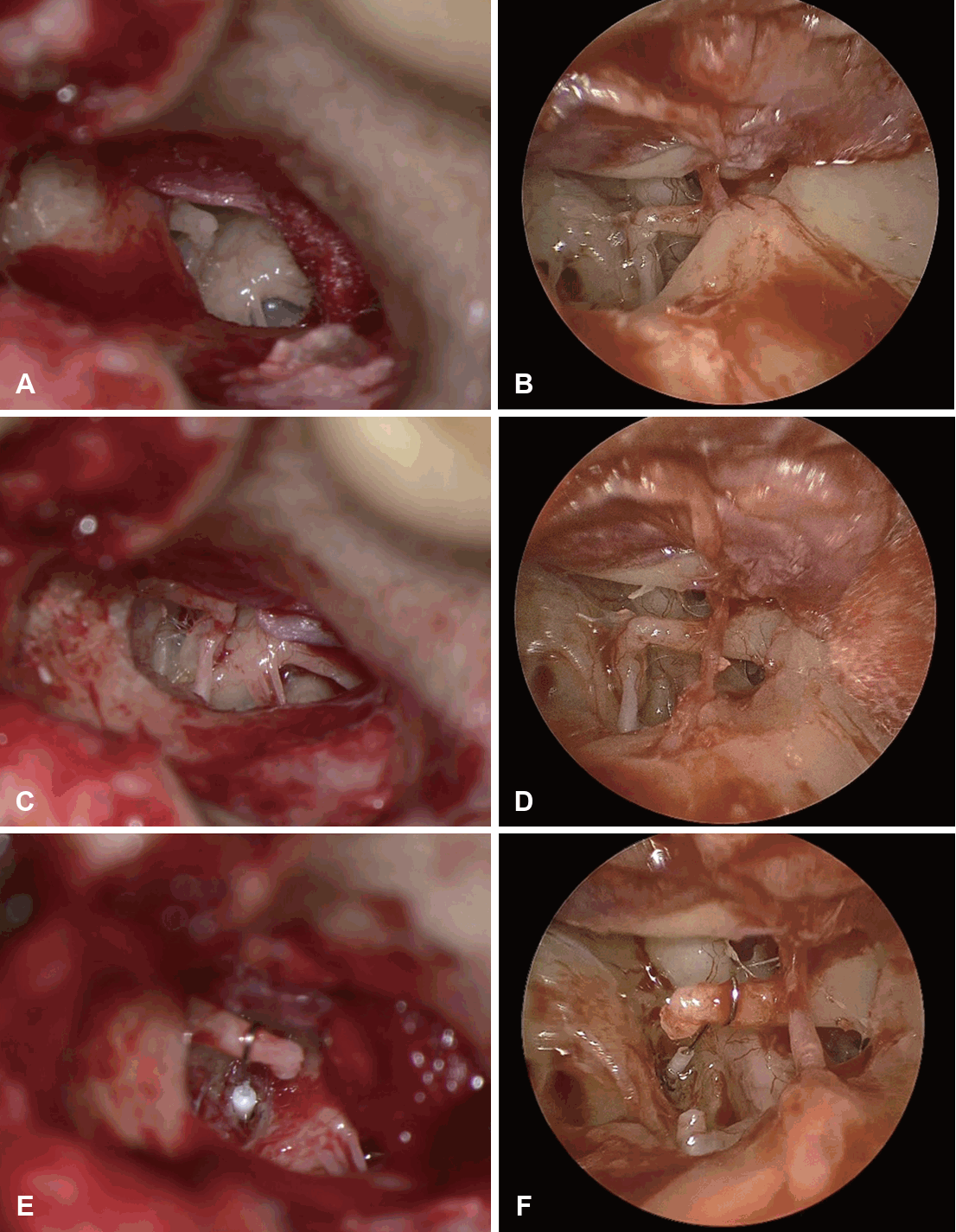
The endoscope provides a wide-angle magnified view of the middle ear cavity. This feature gives ESS a superior field of view compared to conventional microscopic stapes surgery (MSS) at key steps in the procedure, with minimal removal of the posterosuperior bony canal wall. When the middle ear cavity is exposed after the elevation of the tympanomeatal flap, only the incudostapedial joint is usually visible using a microscopic approach (Fig. 2A). In contrast, the endoscopic approach (Fig. 2B) can visualize not only the incudostapedial joint but also the stapes suprastructure and stapes tendon. Unlike the microscopic approach (Fig. 2C), when the posterosuperior bony canal wall is partially removed, the endoscopic approach can visualize the round window inferiorly and the tympanic segment of the facial nerve superiorly in a single field of view (Fig. 2D). In addition, the malleus handle can be observed anteriorly, which can be helpful if the prosthesis needs to be anchored to the malleus head during malleostapedotomy. Moreover, once the prosthesis is placed, the endoscopic approach provides a panoramic view of the adjacent structures, which is advantageous for accurate prosthesis positioning (Fig. 2E and F) and is not possible using the microscopic approach. These visualization advantages can be of great benefit in cases where the stapes and/or other ossicular malformations are present simultaneously with stapes footplate fixation [7].

Comparison of visibility between microscopic and endoscopic stapes surgery. A and B: After tympanomeatal flap elevation. C and D: After partial removal of the posterosuperior bony canal wall. E and F: After placement of the prosthesis.
Despite these advantages, there are also several disadvantages. The endoscopic field of view is a two-dimensional image obtained monocularly and lacks depth perception [8]. This lack of perspective can be overcome by gaining anatomical knowledge of the middle ear structures and by repeatedly moving the scope medially and laterally during surgery. Recently, several techniques have been investigated to improve intraoperative perspective and stereoscopic sensation using computer-based three-dimensional endoscopic systems [8-10]. ESS also carries the risk of causing thermal injury to the inner ear due to heat originating from the light source through the stapedotomy fenestra [11]. Therefore, it is recommended to set the brightness of the endoscopic light source to less than 50% and use suction to prevent the temperature from rising excessively [12]. However, the microscopic approach allows the surgeon to use both hands to manipulate the instruments required for surgery, while the endoscopic approach has the disadvantage of being a one-handed surgical technique. Particularly, the one-handed nature of the procedure makes prosthesis anchoring challenging. The use of a bucket handle prosthesis or self-crimping nitinol prosthesis can reduce this difficulty by eliminating the need for manual crimping using a crimper or alligator forceps [13].
Surgical Outcomes
The postoperative hearing outcomes of ESS are comparable with those of MSS. In a recent pooled analysis of 259 ESS cases in 10 retrospective studies, closure of the air-bone gap within 10 dB was achieved in 71% of cases and within 20 dB in 97% [14]. In randomized controlled studies comparing the outcomes of ESS and MSS, no difference in hearing outcomes was found between the two approaches [15,16].
The incidence of chorda tympani nerve injury is significantly lower with ESS compared to MSS. In a meta-analysis of six studies comparing ESS and MSS, chorda tympani injury was more than three times higher in MSS than in ESS [17]. This lower incidence of chorda tympani injury in ESS may be due to the reduced removal of the posterosuperior bony canal wall [14]. However, regardless of chorda tympani transection, postoperative dysgeusia may occur in 5%-10% of cases because of manipulation of the chorda tympani, such as repositioning or traction, rather than transection [14,18]. To reduce nerve damage, one study suggested a 30-degree endoscopic pre-chorda tympani approach, where a prosthesis is inserted anterior to the nerve, unless the intratympanic portion is of the detached or attached long type [19].
Tympanic membrane perforation can occur postoperatively. A pooled analysis reported an incidence of tympanic membrane perforation of approximately 4.2%, which is higher than the incidence of MSS reported in the literature (0.2%-0.7%) [14]. This high rate of tympanic membrane perforation is thought to be due to the steep learning curve of ESS, which has a significant impact on operative time. Iannella and Magliulo [20] reported the results of 20 ESSs and observed a significant reduction in operative time in the latter 10 cases compared to the first 10 cases, indicating the existence of a steep learning curve for ESS. A recent study analyzing the results of ESS over a 10-year period also showed a decrease in the operative time with accumulating experience [21].
Conclusion
Endoscopic visibility during ESS not only provides a closeup view of the stapes but also reduces injury to the chorda tympani nerve. Moreover, the postoperative hearing results of ESS are comparable to those of MSS. However, prosthesis insertion may be more difficult with ESS than MSS. In addition, the learning curve can lead to longer operation times and a higher incidence of complications, such as perforation of the tympanic membrane during the initial operations. If the surgeon is properly trained using a simulator and/or modular trainer and considers these advantages and disadvantages with a thorough knowledge of middle ear anatomy, ESS can be an alternative to conventional MSS.
Notes
Acknowledgments
The Institutional Review Board and Hospital Research Ethics Committee of Chungnam National University Hospital (Daejeon, Korea) approved this study (CNUH-25-0129).
Author Contribution
Conceptualization: Seongjun Moon, Jin Woong Choi. Data curation: Seongjun Moon. Formal analysis: Seongjun Moon. Funding acquisition: Jin Woong Choi. Methodology: Seongjun Moon. Writing—original draft: Seongjun Moon. Writing—review & editing: Jin Woong Choi.
