Reconstruction of Defects in the Concha and Antihelix Following Oncologic Resection Using a Revolving Door Flap
이개 중앙부 복합 결손 재건을 위한 후이개 아일랜드 피판의 유용성
Article information

Abstract
Reconstruction of conchal and antihelical defects following oncologic resection is challenging due to the complex three-dimensional anatomy and limited local tissue. We report a case of recurrent basal cell carcinoma involving the cavum concha and antihelix in a 75-year-old man. Following complete excision with Mohs micrographic surgery, a significant full-thickness defect was created. A postauricular island flap (revolving door flap) based on the posterior auricular artery was elevated and transposed anteriorly through a cartilaginous window. Unlike skin grafts, this flap provided sufficient soft tissue volume to compensate for the lost cartilage, restoring the natural auricular contour and preventing depression deformity. The postoperative course was uneventful with no flap necrosis. At 1-year follow-up, the patient demonstrated excellent aesthetic outcomes with preserved external auditory canal patency and no tumor recurrence. The revolving door flap is a reliable option for reconstructing complex auricular defects, offering superior vascularity and structural stability.
Introduction
The auricle is part of the auditory apparatus and consists of a complex three-dimensional cartilaginous framework. Because it is externally exposed, it serves important functional and aesthetic roles [1]. Various masses can arise in the auricle. These range from benign lesions such as keloids and hemangiomas to malignant tumors such as basal cell carcinoma, squamous cell carcinoma, and malignant melanoma, the latter of which are commonly documented in the literature [2].
When malignant tumors of the auricle are surgically excised, securing an adequate safety margin is essential. However, the auricle has limited subcutaneous tissue, and the skin is firmly adherent to the underlying cartilage. Therefore, primary closure of defects after wide excision is difficult and may easily result in auricular deformity. Accordingly, selecting an appropriate reconstructive method based on the defect’s location and extent is crucial.
According to Weerda’s classification, defects of the central auricle, including the concha and antihelix, are difficult sites in which to recreate the structural three-dimensional contour [3]. In particular, defects accompanied by cartilage loss carry a high risk of depressed deformity or contracture after simple skin grafting. We successfully reconstructed a composite defect after excision of basal cell carcinoma in the central auricle using a posterior auricular island flap, also known as a revolving door flap, based on the dermal vascular pedicle of the posterior auricular artery. Here, we report this case with a review of the literature.
Case
A 75-year-old man was referred to our department after biopsy confirmed recurrence of basal cell carcinoma of the right concha. He had undergone four previous operations for basal cell carcinoma at the same site and had recently developed pruritus and discharge. On initial examination, an eczematous lesion was observed in the right concha, extending from the antihelix to the entrance of the cartilaginous portion of the external auditory canal (Fig. 1).
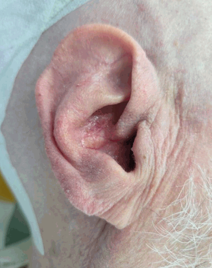
Preoperative local findings. An erythematous, eczematous lesion consistent with recurrent basal cell carcinoma is observed in the right concha and antihelix. A 3 mm safety margin from the tumor border was marked for Mohs micrographic surgery.
In consultation with a dermatologist, we planned Mohs micrographic surgery. We performed the excision with a 3 mm safety margin from the tumor border. Frozen-section examination revealed residual tumor at the external auditory canalside margin, and we excised an additional 5 mm. After confirming that all margins were negative, we completed the resection. The resulting auricular defect was extensive, extending superiorly to the inferior crus of the antihelix, inferiorly to the intertragal incisura, anteriorly to the osseocartilaginous junction of the external auditory canal, and posteriorly to the antihelix. Because tumor invasion was suspected, the defect was full thickness and included the underlying auricular cartilage (Fig. 2).
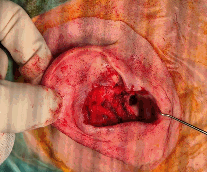
Intraoperative findings. After complete excision of the recurrent basal cell carcinoma, an extensive full-thickness defect involving the right concha and antihelix is observed.
After trimming the margins of the remaining cartilage around the defect, reconstruction was performed using only a soft-tissue flap, without additional cartilage grafting. A postauricular island flap was planned for reconstruction (Fig. 3A). A flap of the same size as the anterior defect was designed on the posterior auricular skin (Fig. 3B). Following careful elevation of the flap while preserving the vascular pedicle containing the dermis and subcutaneous tissue (Fig. 3C), the flap was transposed to the anterior surface through a cartilage window, the defect was reconstructed, and the donor site was primarily closed (Fig. 3D). No postoperative complications, such as auricular deformity or flap necrosis, occurred, and the functional and aesthetic outcomes were favorable. At 1-year postoperative follow-up, there was no evidence of tumor recurrence (Fig. 4). Given his history of repeated recurrence of basal cell carcinoma at the same site, long-term outpatient follow-up will be continued.
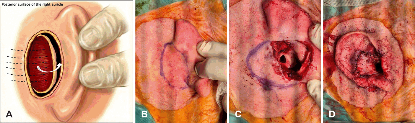
Surgical technique using a postauricular island flap, also known as a revolving door flap. A: Schematic illustration showing transposition of the postauricular island flap through a cartilage window into the anterior defect. B: Flap design on the posterior auricular surface, matched to the size of the anterior defect. C: Flap elevation after postauricular incision while preserving the blood supply from the posterior auricular artery. D: Immediate postoperative appearance after transposition of the flap to the anterior surface, reconstruction of the defect, and primary closure of the donor site.
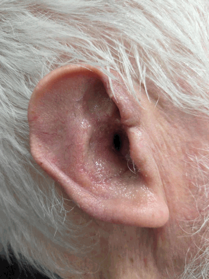
One-year postoperative findings. The reconstructed auricle maintains a natural contour and stable soft-tissue volume, with no secondary depression deformity. The external auditory canal is well preserved, and the postauricular donor-site scar is inconspicuous. No evidence of local recurrence is observed.
This study was approved by the Institutional Review Board of Pusan National University Hospital (IRB No. 2602-017-160). The study was conducted in accordance with the principles of the Declaration of Helsinki.
Discussion
Although the auricle is not located at the center of the face, its distinctive shape and symmetry have a substantial effect on patients’ self-perception and well-being [4]. Therefore, complete excision remains the primary goal in tumor removal, but surgeons must also consider aesthetic reconstruction based on the anatomical characteristics of the auricle. Approximately 8%-10% of all skin cancers arise in the auricle, and basal cell carcinoma is the most commonly reported type [5]. Reconstructing defects after excision of malignant tumors with an adequate safety margin presents a considerable surgical challenge. This challenge stems from the auricle’s unique anatomy: limited subcutaneous tissue, firm adherence of the skin to the cartilage, a restricted local tissue reservoir, and a complex three-dimensional structure in which concave and convex surfaces intersect. In particular, auricular skin has limited intrinsic skin mobility, and primary closure under excessive tension may lead to complications such as skin necrosis or cartilage exposure. Brodland emphasized that, in conchal defects, the mobility of surrounding tissue is extremely limited, making it essential to select an appropriate reconstructive technique according to the size and depth of the defect rather than relying on simple closure [6].
Skin grafting is one of the most commonly considered options for reconstruction of central auricular defects and has the advantage of technical simplicity. However, it also has several limitations. Talmi, et al. [7] noted that when skin grafting is performed for anterior auricular defects, the absence of an underlying supporting structure may result in depression deformity, contracture, and color mismatch with surrounding tissue. In addition, graft take rates are low in areas of exposed cartilage, leaving a persistent risk of necrosis.
The postauricular island flap used in the present case can effectively overcome these limitations. This technique, first described by Masson [8], provides abundant blood supply through a dermal pedicle and therefore allows a high survival rate regardless of cartilage exposure. Dessy, et al. [9] reported that this flap provides sufficient soft tissue volume, compensates for the absence of the removed cartilage, and is highly effective for restoring the natural three-dimensional contour of the auricle. In the present case, follow-up at 2 weeks and 6 months confirmed maintenance of a stable three-dimensional contour without the depression commonly observed after skin grafting.
Other options for reconstruction of central auricular defects include preauricular flaps and perichondrial-cutaneous composite grafts. However, preauricular flaps may leave visible facial scars, and composite grafts may show less reliable graft survival when the defect is large. In contrast, the postauricular island flap conceals the donor-site scar within the postauricular sulcus and provides color and texture that closely match those of the defect.
A potential drawback of this technique is reduced auricular prominence, particularly when it is used for large defects. During transposition of posterior auricular tissue to the anterior surface, tension from donor-site closure may reduce the auriculocephalic angle, which can lead to decreased postoperative auricular prominence. Therefore, surgeons should carefully consider donor-site laxity during flap design and explain preoperatively that symmetry between the two ears may be reduced. A more fundamental limitation of this technique is encountered when the auricular defect is so extensive that the overall supporting framework of the auricle has completely collapsed. In such large defects, reconstruction using only a soft-tissue flap is limited, and framework reconstruction using costal cartilage may produce better results for reproducing the delicate three-dimensional structure of the ear [10]. However, costal cartilage reconstruction involves more extensive surgery and carries a risk of donor-site complications. Therefore, in composite defects confined to the central auricle, as in the present case, the postauricular island flap can serve as a less invasive yet effective reconstructive option.
This technique can be safely applied not only to defects confined to the concha but also to extensive central composite defects involving the antihelix or scapha. However, the present case reports outcomes only up to 1 year after surgery. Given the patient’s history of repeated recurrence of basal cell carcinoma at the same site, longer-term follow-up is required. We plan to continue regular outpatient follow-up to evaluate both tumor recurrence and the morphological stability of the reconstructed auricle. In conclusion, reconstruction using a postauricular island flap is an effective and reliable method for auricular defects after excision of malignant tumors in the central auricle, while achieving both functional preservation and aesthetic reconstruction.
Supplementary Materials
Korean translation of this article is available with the Online-only Data Supplement at https://doi.org/10.3342/kjorl-hns.2026.00115.
Notes
Acknowledgments
This work was supported by clinical research grant from Pusan National University Hospital in 2026.
Author Contribution
Conceptualization: Se-Joon Oh. Data curation: Han-Seul Na. Formal analysis: Han-Seul Na. Investigation: Sung-Won Choi, Hoon-Soo Kim, Se-Joon Oh. Methodology: Hoon-Soo Kim, Se-Joon Oh. Project administration: Se-Joon Oh. Resources: Hoon-Soo Kim. Supervision: Sung-Won Choi, Se-Joon Oh. Validation: Sung-Won Choi, Hoon-Soo Kim. Visualization: Han-Seul Na, Hoon-Soo Kim. Writing—original draft: Han-Seul Na. Writing—review & editing: Han-Seul Na, Se-Joon Oh.