A Case of Relapsing Polychondritis Presenting as a Bilateral Sequential Perilymphatic Fistula
양측에 순차적으로 발생한 외림프 누공으로 발현된 재발성 다발 연골염 1예
Article information

Abstract
Relapsing polychondritis (RPC) is a rare systemic autoinflammatory disease that primarily affects cartilaginous organs. Inner ear involvement occurs in up to 40% of patients, leading to fluctuating or sudden audiovestibular dysfunction. The proposed mechanisms include autoimmune reactions to the membranous labyrinth and immune-mediated vasculitis. We report the case of a 21-year-old man with RPC who presented with sequential bilateral perilymphatic fistula (PLF) and acute audiovestibular loss. He first experienced aggravated vertigo and fluctuating hearing loss (HL) on the left side 5 days before the visit. Exploratory tympanotomy for the suspected PLF produced immediate improvement. Twenty days later, he experienced sudden vertigo, right-sided HL, and bilateral auricular swelling. CT revealed soft-tissue density in the right round-window niche, and perilymph leakage was confirmed surgically. Bilateral auricular chondritis and episcleritis prompted the diagnosis of RPC. This case suggests that PLF may represent an additional mechanism of rapid audiovestibular loss in RPC and that prompt surgical repair with immunosuppression may preserve hearing.
Introduction
Relapsing polychondritis (RPC) is a clinically fluctuating, progressive condition characterized by recurrent episodes of inflammation involving cartilaginous organs, resulting in anatomical deformities and functional impairment of the affected structures. All types of cartilage may be affected, including the elastic cartilage of the ears and nose, hyaline cartilage of peripheral joints, fibrocartilage of the axial skeleton, and cartilage of the tracheobronchial tree. The pathology may also involve other proteoglycan-rich structures, such as the conjunctiva and sclera of the eye, media of the arteries, and inner ear [1-3].
Involvement of the inner ear in RPC manifests as audiovestibular loss, such as hearing loss (HL), tinnitus, and vertigo, which accounts for 13% to 53% of cases [4,5]. The presentation of these inner ear symptoms can vary from being slowly progressive over several years to being rapidly progressive, resulting in profound bilateral audiovestibular loss. The pathomechanism of audiovestibular symptoms has been attributed to autoimmune inflammation of inner ear structures. Here, we report a case of RPC presenting with a bilateral perilymphatic fistula (PLF), which developed sequentially from episcleritis to bilateral PLF and subsequently to bilateral auricular perichondritis. This case suggests that RPC-related autoimmune reaction may be implicated in the fragility of the oval and round windows (RWs), which could potentially lead to PLF as an additional mechanism for audiovestibular loss.
Case
A 21-year-old man presented to the emergency center with sudden aggravated vertigo and fluctuating ear fullness/tinnitus in his left ear. He had experienced dizziness 1 month prior after being hit by a soccer ball, which improved at that time but worsened again 5 days before the visit. He had recently experienced recurrent red eye for 3 months that improved with repeated oral administration of low-dose steroids prescribed at a local ophthalmologic clinic but had been otherwise healthy. Although both eardrums appeared normal, he showed mixed-type HL in the left ear and right-beating spontaneous nystagmus augmented by head shaking. Typical left vestibular hypofunction was recorded, with 80% canal paresis on bithermal caloric test and a 71% reduction in the cervical vestibular evoked myogenic potential response. Traumatic PLF was suspected, and a high-dose steroid was administered with absolute bed rest. Nevertheless, dizziness continued, and the hearing threshold worsened (Fig. 1). Although temporal bone computed tomography (TBCT) revealed negative findings (Fig. 2B), exploratory tympanotomy of the left ear was performed on day 5. Although a PLF was not clearly visualized intraoperatively, the surrounding mucosa was refreshed and both round and oval window areas were tightly packed with pieces of connective tissue and fibrin glue. His vertigo symptoms and bone conduction thresholds improved postoperatively, and he was discharged from the hospital on postoperative day 4. Two weeks later, he returned with left auricular swelling and otalgia. Perichondritis was diagnosed, and antibiotics were prescribed.
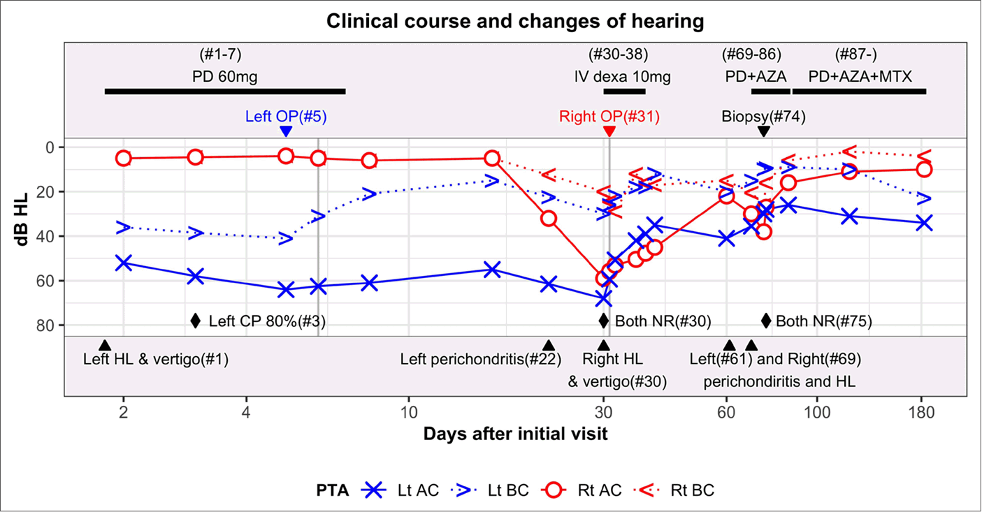
Clinical course and changes in hearing thresholds in the patient. The timeline illustrates the rapid, step-by-step progression characteristic of this case: beginning with the initial ocular symptoms, followed by left-sided audiovestibular loss requiring surgery, subsequent auricular chondritis, and the consecutive development of right-sided audiovestibular loss, which ultimately led to the rheumatologic and ophthalmologic confirmation of relapsing polychondritis. The x-axis represents the days elapsed since the initial visit. The upper panel describes the medical treatments (horizontal black bar) and surgical interventions (lower arrowhead) provided, and the lower panel denotes the days of symptom development (upper arrowhead). Changes in the hearing threshold are plotted in the middle panel according to the y-axis. The diamonds mark the timing of the bithermal caloric tests and results. HL, hearing loss; PTA, pure tone audiometry; AC, air conduction; BC, bone conduction; OP, operation; PD, prednisolone; AZA, azathioprine; MTX, methotrexate; CP, canal paresis; NR, no response.
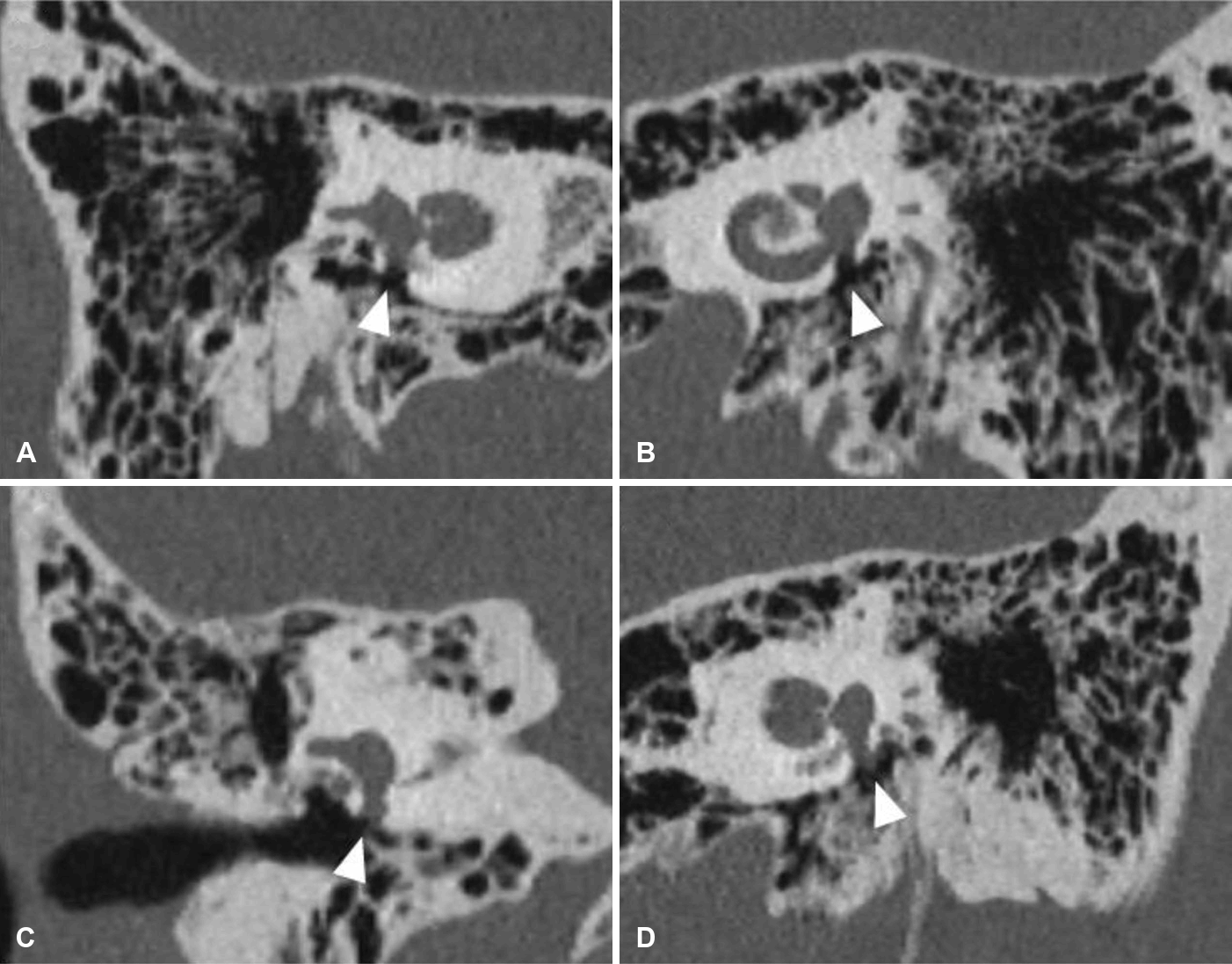
TBCT findings of the patient. The round window area that showed air on the initial TBCT (taken on day 1; A and B) shows soft-tissue density on the second TBCT (taken on day 30), which suggests perilymphatic leakage in the right ear (C) and postoperative change in the left ear (D). TBCT, temporal bone computed tomography.
On day 30, he returned to the emergency center with sudden right-sided HL and vertigo without improvement of perichondritis in the left ear. Mixed-type HL in the right ear and left-beating nystagmus were noted. The bithermal caloric test revealed bilateral vestibular hypofunction, confirmed by an ice-water test, revealing the observed spontaneous nystagmus as the Bechterew phenomenon [6]. Repeated TBCT revealed soft-tissue density at the round and oval window areas of the right ear (Fig. 2C). After the administration of a high-dose steroid, an exploratory tympanotomy of the right ear was performed the following day. Direct perilymph leakage was observed through the RWs (S1), and subsequently, both round / oval windows were tightly sealed with pieces of connective tissue. The audiovestibular symptoms and left perichondritis improved postoperatively. One month after the 2nd operation, bilateral auricular swelling developed sequentially (Fig. 1, days 61 and 69), accompanied by a 10-20 dB hearing decline in both ears. Immunosuppressive medication was initiated after consultation with a rheumatologist under the suspicion of RPC, which was later supported by histopathological findings (Fig. 3). On consultation with an ophthalmologist, episcleritis was diagnosed such that the previous steroid-responsive ocular symptoms were the first presenting symptoms of RPC in this case. Since then, the patient has undergone follow-up examinations by a rheumatologist and physicians in our department with stable bilateral hearing while maintaining immunosuppressive treatment and vestibular rehabilitation exercise. This case report was exempted by the Institutional Review Board of Hallym University Sacred Heart Hospital (HALLYM 2021-05-012).
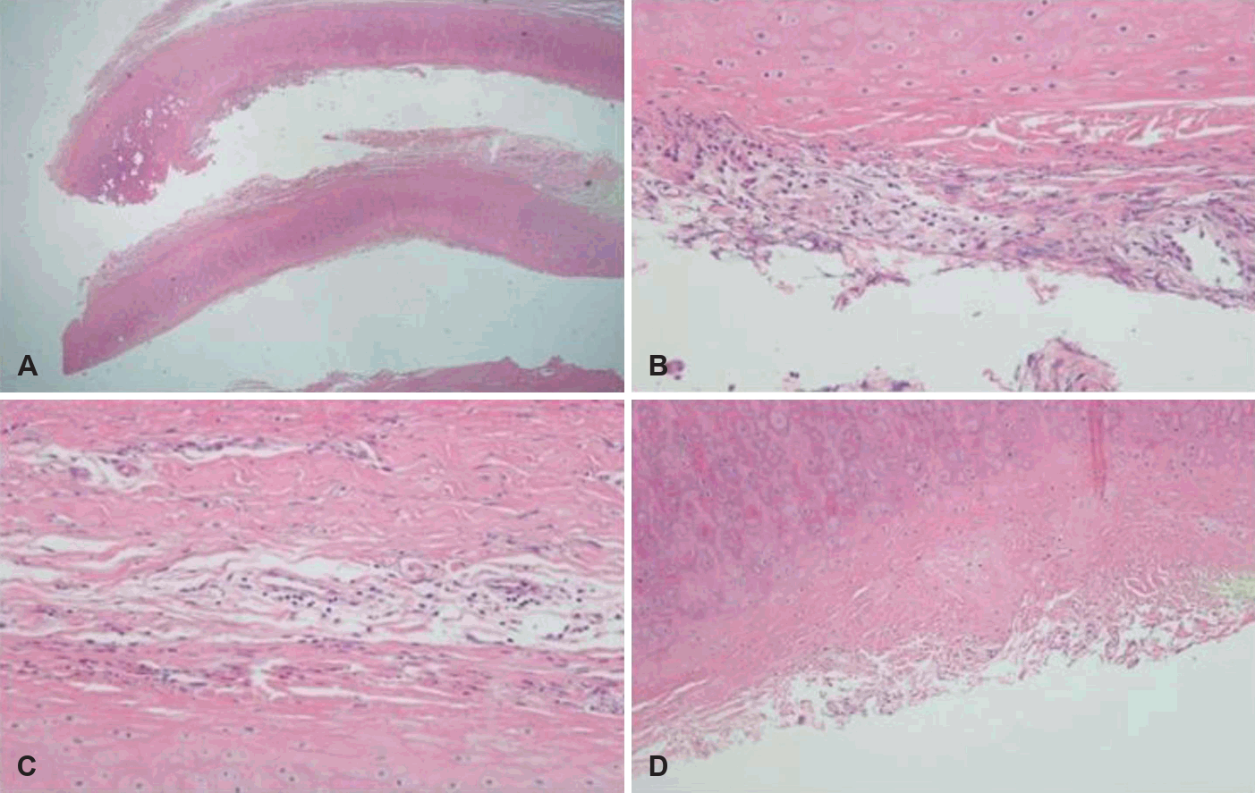
Histologic findings of the auricular cartilage (H&E staining). (A) Low-power view shows the general layered architecture of the auricular cartilage. Higher magnifications demonstrate (B and C) mild lymphocyte and mononuclear cell infiltration within the perichondrium, along with (D) fibrotic changes causing irregular margins. These findings, lacking the dense neutrophilic exudate or abscess formation typical of acute infectious perichondritis, support the histopathological diagnosis of relapsing polychondritis.
Discussion
This study reports a case of RPC with bilateral sequential PLF as a presenting symptom. The patient met the diagnostic criteria for probable PLF in the left ear—a preceding barotraumatic event, sudden auditory symptoms, and dizziness with positional characteristics—and was diagnosed with definite PLF in the right ear based on visual confirmation of perilymph leakage during surgery. Ultimately, the patient was diagnosed with RPC accompanied by bilateral perichondritis and ophthalmologic manifestations [7,8]. With surgical intervention, the patient could be maintained at a mild degree of HL or better in both ears.
The diagnosis of RPC is based on clinical symptoms, with no definitive laboratory markers. According to McAdam’s criteria, three or more of the following six clinical findings are required: 1) bilateral auricular chondritis, 2) polyarthritis, 3) nasal chondritis, 4) ocular inflammation, 5) respiratory tract chondritis, and 6) audiovestibular damage. Cartilage biopsy can sometimes be applied in cases of diagnostic uncertainty. Given the history of blunt head trauma, this case was initially suspected of having barotraumatic PLF. While RPC is generally known to have a slow and progressive course, the abrupt and sequential development of symptoms in this patient might represent an atypical, rapid flare-up. Therefore, we hypothesize that physical trauma may have acted as an aggravating factor, potentially accelerating the development of PLF associated with underlying RPC-related autoimmune inflammation. The abrupt and sequential development of ear symptoms prompted surgical intervention to stop progression of HL. After surgical repair of the bilateral ears, the patient subsequently developed bilateral auricular chondritis, and a recent history of ocular inflammation confirmed the diagnosis of RPC.
The pathogenesis of RPC is mediated by autoimmune reactions to type II collagen, which is abundant in cartilage and widely distributed in the inner ear, including the round/oval window area [9]. In addition, the pathomechanism of inner ear disorders has been considered an autoimmune reaction to the membranous labyrinth or vasculitis [10]. The inner ear symptoms caused by the above pathomechanism mainly consist of progressive sensorineural HL that is partially responsive to steroids [11]. However, as in this case, rapid progression of HL leading to a profound degree of vestibular areflexia could occur in RPC. In this case of RPC, in which the patient presented with preceding inner ear symptoms, bilateral PLF was diagnosed based on the clinical response to surgery in the left ear and surgical confirmation in the right ear, suggesting that the rapid progression of HL in RPC can be better explained by PLF than by the previously proposed mechanisms.
Whether PLF developed idiopathically or secondary to RPC remains uncertain. The RW membrane consists of three layers: the tympanic cavity layer, the middle connective tissue layer, and the scala tympani layer [12]. The middle connective tissue layer, which is considered the most important part of the RW membrane because it permits the movements of the inner ear fluid caused by the movements of the stapedial footplate, consists of collagen and elastic fibers [13]. Because collagen is present in the RW area, an autoimmune reaction in these areas of RPC could lead to PLF, leading to profound audiovestibular loss.
Unlike HL caused by autoimmunity of the collagen structure of the inner ear, which often improves with steroids, acute profound HL manifesting as PLF may require immediate surgical intervention. In this case, HL improved in the previously unoperated left ear after the second surgery and improved in both ears following immunosuppressive treatment after the diagnosis of RPC. Therefore, in this case, it can be inferred that HL was likely accompanied by autoimmune involvement of the inner ear structures as well as PLF. However, prompt surgical management appeared to improve pseudoconductive HL during sudden attacks, allowing the patient to maintain normal to mild hearing levels in both ears. Specifically, the mixed HL observed can be attributed to a third-window phenomenon created by the PLF, which may manifest as pseudoconductive HL [14]. Although surgical sealing of the fistula improved bone conduction, the air-bone gap might not have sufficiently decreased immediately due to postoperative factors such as middle ear packing or transient effusion. The subsequent improvement in air conduction observed during follow-up was likely achieved as the packing materials and surgical glue were gradually absorbed over time.
This case suggests that PLF could be a possible mechanism of inner ear symptoms in RPC and that immediate intervention may contribute to preserving audiovestibular function.
Supplementary Materials
The Supplement is available with this article at https://doi.org/10.3342/kjorl-hns.2026.00136.
Notes
Acknowledgments
This study was supported by the Basic Science Research Program through the National Research Foundation of Korea (NRF), funded by the Ministry of Science and ICT [grant number RS-2022-NR069203].
Author Contribution
Conceptualization: Hyo-Jeong Lee. Data curation: Doo Sik Park, Soomin Jo. Formal analysis: Soomin Jo. Funding acquisition: Hyo-Jeong Lee. Investigation: Doo Sik Park, Haewon Rhi. Methodology: Haewon Rhi. Project administration: Soomin Jo. Resources: Hyo-Jeong Lee. Software: Hyo-Jeong Lee. Supervision: Hyo-Jeong Lee. Validation: Soomin Jo. Visualization: Haewon Rhi. Writing—original draft: Doo Sik Park, Soomin Jo. Writing—review & editing: all authors.