Extended Columellar Strut Using an Integrated Cartilage-Bone Composite Graft: A Technical Description
연장 비주 지지대로서의 맞춤형 연골-골 복합 이식물
Article information

Abstract
Achieving stable nasal tip projection in Asian rhinoplasty is frequently limited by insufficient septal cartilage, particularly in cases of age-related ossification or post-traumatic septal change. We describe an endonasal technique in which the posterior nasal septum is harvested as a single integrated cartilage-bone unit, preserving the native bone-cartilage junction without separation, and used as an extended columellar strut for tip augmentation. The integrated bony component provides structural rigidity that pure cartilage alone may not offer, reducing the need for supplementary cap grafting and preserving a natural infratip lobule-to-nostril proportion. Candidate selection is guided by preoperative CT assessment of bony thickness and junction integrity at the planned harvest site. Key technical principles include controlled incremental drilling, precise pocket dissection, and avoidance of tensioned fixation to the lower lateral cartilages. We present an illustrative case of a 40-year-old male with post-traumatic partial septal ossification and low nasal tip deformity; we applied this technique and observed satisfactory aesthetic outcome at the 3-month follow-up. This report describes the operative concept and technical approach; however, it does not include outcome data nor does it claim comparative superiority.
Introduction
In Asian populations, augmentation rhinoplasty significantly predominates over reduction procedures [1,2]. While alloplastic materials such as silicone or expanded polytetrafluoroethylene are routinely used for dorsal or radix augmentation, autografts remain strongly preferred for nasal tip surgery to minimize the risks of extrusion, infection, and long-term complications.
Achieving adequate tip projection in Asian patients, however, presents consistent challenges. A substantial proportion of patients present with a short nose deformity [2], and the available nasal septal cartilage is often reduced due to age-related ossification or post-traumatic calcification of the posterior septum [3]. When pure cartilage is insufficient, costal cartilage represents a well-established alternative; however, its use carries recognized drawbacks including increased operative time and costs, potential donor-site morbidity, and the risk of postoperative warping [4].
In this article, we describe a practical endonasal technique in which the posterior nasal septum is harvested as an integrated cartilage-bone composite unit, without separating the bony and cartilaginous components. This approach provides a single-unit graft with inherent structural rigidity, sufficient length for extended columellar strut (ECS) placement, and avoids the reassembly steps required in previously described composite techniques. We present the technical steps, operative pearls, limitations, and an illustrative case.
Indications and Patient Selection
This technique is indicated in the following clinical scenarios:
• Patients with a low nasal tip or short nose deformity requiring substantial tip projection, in whom the available pure septal cartilage is insufficient in either length or structural integrity after preserving an adequate L-strut.
• Patients in whom septal cartilage length is adequate but thickness is insufficient to provide the structural rigidity required for a stable ECS.
• Patients with a history of nasal trauma, prior nasal surgery, or advanced age in whom partial or complete ossification of the posterior nasal septum is anticipated or confirmed on preoperative CT imaging, limiting the availability of pure cartilage.
• Patients who prefer a nasal tip that retains a degree of natural postoperative elasticity rather than a rigidly fixed tip.
The technique is not intended as the primary approach in patients with adequate and healthy septal cartilage. In those cases, standard ECS grafting with pure septal cartilage remains preferable. The following factors should be considered relative contraindications or situations where alternative donor sites should be planned:
• Severely calcified or completely ossified posterior septum with a fragile or irregular bony architecture, in which controlled drilling is not technically feasible or the risk of graft fracture is high.
• A posterior bony septum that is judged on preoperative CT to be too thin to yield a graft of sufficient structural rigidity for columellar strut use. When significant doubt exists, alternative donor sites should be considered preoperatively.
• Cases requiring major structural reconstruction of the nasal framework, extensive spreader grafting for valve collapse, or complex revision rhinoplasty with significant dorsal or caudal septal deficiency.
• Patients with a preexisting upturned nasal tip (excessive cephalic tip rotation) or those in whom correction of tip rotation is the primary surgical objective.
Preoperative Evaluation
Clinical photography
Standardized facial photographs including frontal, profile, oblique, and basal views are obtained. These establish precise documentation and facilitate surgical planning for rhinoplasty.
CT assessment
Preoperative CT imaging of the nasal septum is essential and is performed in all candidates for this technique. The midsagittal and coronal CT views are reviewed to:
• Determine the extent of the L-strut that must be preserved (a minimum of 10-15 mm of caudal and dorsal strut is maintained).
• Assess the quantity and quality of harvestable septal cartilage posterior to the L-strut.
• Evaluate the posterior septal bone, including the perpendicular plate of the ethmoid (PPE) and vomer, for thickness, regularity, and the presence of partial ossification at the cartilage-bone junction (Fig. 1).
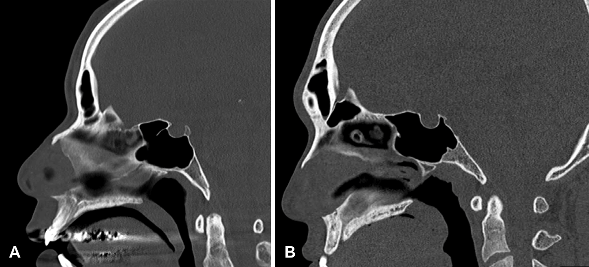
Preoperative CT assessment of the nasal septum. A: Older patient with partial ossification: midsagittal CT view demonstrates progressive ossification of the posterior septal cartilage. The harvestable area of pure cartilage (after preserving the L-strut) is limited. B: Young patient in the third decade: the septal cartilage shows minimal ossification, providing a generous supply of pure cartilage.
• Estimate the achievable graft length and width, and identify the optimal harvest zone.
The upper portion of the PPE tends to be thicker and more suitable for drilling, whereas the lower portion is thinner and more fragile. The posterior vomer is generally thick and may provide supplementary bony support.
Surgical Technique
Step 1: harvest of cartilage-bone composite graft via septoplasty
Incision and flap elevation
A modified Killian incision is placed approximately 5 mm posterior to the mucocutaneous junction. Meticulous bilateral subperichondrial and subperiosteal flap elevation is performed to expose the septal cartilage and the posterior bony septum.
L-strut preservation
An L-strut of at least 10-15 mm in both the dorsal and caudal dimensions is strictly maintained to preserve adequate structural support of the nasal framework.
Graft harvesting
An incision is made through the septal cartilage with a Dknife, posterior to the L-strut. Crucially, the posterior bone is not separated from the cartilage at this stage. Instead, a surgical drill (preferably a high-speed drill with appropriate irrigation) is used to cut through the bony septum along the planned margins (Fig. 2C). Drilling is applied gradually and evenly to avoid abrupt fracture, uncontrolled cracking, or separation of the cartilage-bone junction.
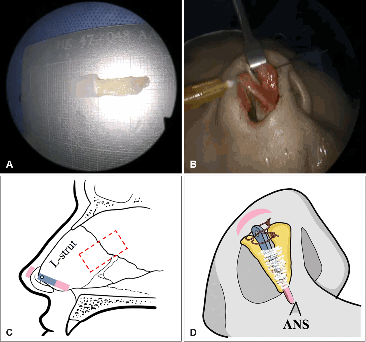
Graft preparation and fixation. A: The harvested cartilage-bone composite graft after back-table preparation, trimmed to approximately 23 mm in length and 3 mm in width. The natural bone-cartilage junction is preserved as a single integrated unit throughout harvesting. B: Intraoperative photograph demonstrating endonasal insertion of the composite graft into the columellar pocket, with the LLCs delivered and prepared for suture fixation. C: Schematic diagram illustrating the harvest design on the nasal septum. The dashed rectangle indicates the planned harvest zone posterior to the preserved L-strut. The blue segment represents the bony component (perpendicular plate of the ethmoid) and the pink segment the cartilaginous component of the L-strut. D: Schematic diagram showing the orientation and fixation of the composite graft within the columella. The cartilaginous end is seated at the anterior nasal spine to minimize mechanical irritation, while the bony end is directed superiorly toward the nasal tip. The dome segments of the LLCs are secured to the bony portion through a pre-drilled fixation hole using 5-0 PDS sutures. LLC, lower lateral cartilage; PDS, Polydioxanone suture.
Drilling technique and depth control
The drill is advanced until the bone is almost completely severed, leaving only a thin bridge. The composite graft is then completed by gently rocking or tapping with a narrow osteotome or Hartmann forceps, producing a controlled fracture. This sequential approach— partial drilling followed by controlled instrumented completion—is key to preventing irregular fracture edges and unintended separation of the bone-cartilage junction.
Graft tailoring
The required graft length is estimated preoperatively from standardized profile photographs, in which the degree of intended tip projection is assessed and used to approximate the necessary strut length. In our experience, a graft length of at least 20 mm is generally required to achieve reliable tip projection and stable seating at the ANS. The graft is trimmed to a width of 2-3 mm to allow secure placement within the dissected pocket while minimizing columellar bulk (Fig. 2A and B). The proportion of bone to cartilage is not predetermined and varies by harvest site. What is essential is that a cartilaginous component is retained, as it provides the flexibility needed for atraumatic pocket insertion and reliable suture fixation to the lower lateral cartilages (LLCs). If the harvested cartilage proves excessively thin or structurally weak on the back table, it should be trimmed back, allowing the bony component to occupy a greater proportion of the graft. A 1 mm diamond-tip drill bit is used to create a small fixation hole in the bony portion to facilitate suture fixation to the LLCs.
Step 2: endonasal rhinoplasty and tip augmentation
Dissection and delivery
Via marginal and transfixion incisions, the LLCs are isolated and delivered. The marginal incision is deliberately extended as far laterally as possible along the lateral crus, allowing maximal mobilization of the LLC. This lateral extension, combined with dissection carried superiorly to the upper lateral cartilages and nasal bones, ensures that the entire LLC can be advanced and secured to the columellar strut without tension, enabling the tip to be elevated to the desired projection while minimizing the risk of cephalic rotation during augmentation.
Medial crura preparation
The LLCs are delivered through a single nostril. The medial crura are partially dissected with converse scissors to create a pocket of approximately 3 mm in width, extending inferiorly to the anterior nasal spine (ANS). Deliberate partial dissection, rather than wide undermining, is preferred: a snugly fitting pocket provides inherent internal resistance that stabilizes the graft in the intended position, whereas excessive dissection creates dead space that permits postoperative graft migration or malposition. The surrounding soft tissue tension acts as a natural retaining force, complementing suture fixation and contributing to long-term positional stability of the strut.
Graft orientation rationale
The composite graft is oriented with the cartilaginous end toward the ANS and the bony end directed superiorly toward the tip (Fig. 2D). We prefer placing the cartilaginous portion at the ANS to enhance patient comfort. Direct contact between the rigid bony graft and the ANS may lead to mechanical irritation and localized pain, particularly during tip manipulation or animation. Positioning the bony segment near the tip also facilitates suture fixation through the pre-drilled hole to the LLCs.
Graft insertion and fixation
The graft is inserted into the pocket and temporarily stabilized with a 27-gauge needle (Fig. 2C). It is then permanently fixed to the LLCs with 5-0 Polydioxanone suture (PDS) at two points, utilizing the pre-drilled hole in the bony portion. Care is taken to ensure that the graft lies along the midline and that fixation is symmetric.
Prevention of graft malposition and bending
Graft bending or dislocation at the bone-cartilage junction remains a theoretical concern under sustained mechanical stress, particularly when excessive tension is placed on the construct during fixation. Specifically, overcorrection of tip projection—achieved by maximally stretching and fixing the LLCs under tension—should be avoided. We recommend that LLC fixation be performed with the tip in a naturally projected, non-tensioned position, allowing the composite strut to provide structural support rather than act as a tension-bearing element.
Final refinement and camouflage
The new tip-columellar complex is repositioned under the skin envelope. Tip projection, definition, and symmetry are assessed and adjusted as needed. A small dermis or cartilage graft may be placed over the tip in a cap fashion if minor surface irregularities are present (Fig. 2C and D).
Additional procedures
Depending on the patient’s unique anatomical needs, dorsal procedures-such as augmentation or osteotomy-can be integrated into the surgical workflow either before or after the tip-plasty.
Technical pearls
• Preoperative CT review is mandatory; the surgeon should identify the specific harvest zone where the bony septum is of adequate thickness and the bone-cartilage junction is intact, and estimate the achievable graft dimensions before making any incision.
• The marginal incision should be extended as far laterally as possible along the lateral crus. Maximal lateral mobilization of the LLC allows the tip to be elevated to the desired projection without tension at the fixation point.
• The drill should be used with controlled, incremental pressure and adequate irrigation to prevent thermal injury and irregular bony fracture.
• The bone-cartilage junction should never be forcibly separated during harvest.
• The pre-drilled fixation hole in the bony portion significantly simplifies secure suture fixation and reduces the risk of suture cut-through during tying.
Pitfalls and contraindications
• Drilling in a completely ossified or very fragile posterior septum may result in irregular fracture edges or inadvertent separation of the bone-cartilage junction, compromising the composite integrity.
• Inadequate L-strut preservation leads to nasal structural collapse; the minimum 10-15 mm L-strut must always be confirmed before harvesting.
• The bony component of the graft may be palpable, particularly in thin-skinned patients; a small dermis graft or soft tissue camouflage can mitigate this.
• This technique does not address complex functional airway pathology requiring spreader grafts for internal valve collapse; alternative or additional grafting strategies should be considered in such cases.
Postoperative Care
Postoperative management follows our standard rhinoplasty protocol. An external nasal splint is applied for 7 days. Internal nasal packing is removed on postoperative day 2. Patients are instructed to avoid nasal trauma and strenuous activity for 4 weeks. Follow-up visits are scheduled at 1 week, 1 month, 3 months, and 6 months postoperatively.
Illustrative Case
The patient was a 40-year-old male presenting with a low nasal tip and short nose deformity. He had a history of nasal bone fracture requiring prior surgical repair. Preoperative profile photographs demonstrated a pseudohump deformity secondary to relative tip underprojection, rather than true dorsal excess (Fig. 2B). The surgical plan therefore included tip projection via an ECS combined with dorsal augmentation to achieve a harmonious nasal profile.
Preoperative CT imaging demonstrated partial ossification of the posterior nasal septum, with the available pure cartilage posterior to the L-strut being insufficient in length for a standard ECS (Fig. 1A). A cartilage-bone composite graft was harvested as a single integrated unit posterior to the L-strut, without separating the cartilaginous and bony components (Fig. 2A and C). The initial harvest was performed as a rectangular block incorporating both the posterior septal cartilage and the adjacent ossified bone. The graft was then refined on the back table using a drill: the bony portion was precisely cut and shaped to yield a final graft of approximately 23 mm in length and 3 mm in width, maintaining a uniform thickness of 1.0-1.5 mm throughout. In this case, given the extent of ossification, the final graft comprised approximately 30% cartilage and 70% bone along its length. A 1 mm fixation hole was pre-drilled in the bony portion.
The endonasal approach was used with marginal and transfixion incisions. Following LLC delivery and pocket preparation, the graft was inserted with the cartilaginous end oriented toward the ANS and fixed to both LLCs with 5-0 PDS sutures through the pre-drilled hole (Fig. 2). A small dermis graft was placed over the tip and dorsum for soft tissue camouflage and to smooth surface irregularities.
Postoperative photographs at 3 months demonstrated clinically meaningful improvement in nasal tip projection and definition, with a natural infratip lobule contour without the bulky appearance associated with multilayer cap grafting (Fig. 3). The patient reported satisfactory aesthetic outcome with no functional complaints. No graft-related complications including palpability, displacement, or asymmetry were observed at follow-up.
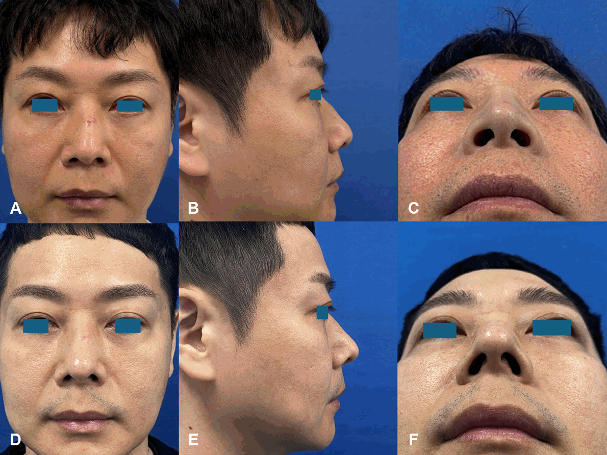
Preoperative and postoperative clinical photographs of the illustrative case. A 40-year-old male with a pseudohump and a low nasal tip deformity. The upper row (A-C) shows preoperative views and the lower row (D-F) shows views at 3 months postoperatively. The pseudohump appearance (B), which was secondary to relative tip underprojection rather than true dorsal excess, has been corrected through tip augmentation combined with modest dorsal refinement, yielding a harmonious nasal profile (E). Basal views. Improved infratip lobule definition is demonstrated without the bulkiness associated with multilayer cap grafting (F). Nostril symmetry is preserved and no columellar irregularity is visible.
This case is illustrative of the technique and its intended application; it is not presented as evidence of routine outcomes across a patient population.
Discussion
The primary challenge in Asian augmentation rhinoplasty is achieving stable and sufficient tip projection when autologous material is limited [1,2]. The progressive ossification of the posterior septal cartilage with age, or following nasal trauma, substantially reduces the harvestable area of viable pure cartilage after preserving an adequate L-strut (Fig. 1) [3]. Costal cartilage is an established alternative in such cases; however, its well-recognized drawbacks—including donor-site morbidity, risk of warping, and increased operative complexity—prompt continued interest in maximizing the utility of available septal material [4]. Given its critical role in complex cases such as revision rhinoplasty, contracted or saddle nose deformities, and reconstructive surgery for congenital or traumatic defects, costal cartilage should be judiciously reserved as a secondary option.
The use of the PPE and septal bone has been previously documented in several applications, including septal extension grafts (SEGs), spreader grafts, and L-strut reinforcement [5-11]. Prior techniques can be broadly categorized as: 1) use of isolated septal bone segments; 2) cartilage-bone complex techniques in which the components are separated and then reassembled, such as the “sandwich technique” described by Ahn, et al. [7] in which ethmoid bone is interposed between cartilage layers; and 3) the “bony cartilaginous unit” concept described by Sazgar [6], applied for caudal extension and lateral crural struts. Wang, et al. [9] described a modified SEG complex combining septal cartilage, ethmoid bone, and auricular cartilage, and Lee, et al. [10] used a septal cartilage-ethmoid bone composite for correction of short nose deformity.
To our knowledge, the use of an integrated septal cartilage-bone composite graft specifically as an ECS has not been previously described. While prior studies have explored cartilage-bone composites, they have primarily focused on SEGs or spreader grafts, or on adding bone segments to reinforce the rigidity of conventional columellar struts. In contrast, our technique utilizes the native, unseparated junction of the bony-cartilaginous unit as a primary structural framework. Among the structural options available, the SEG is a powerful technique capable of achieving substantial tip projection and rotation. However, SEG inherently sacrifices a degree of natural tip mobility and typically requires a larger volume of graft material. It is therefore best reserved for cases of severe short nose deformity or those in which significant cephalic tip rotation must be corrected. For the majority of patients seeking natural-appearing tip augmentation without major rotational change, the ECS offers a more aesthetically refined outcome with less structural rigidity at the tip complex.
When pure septal cartilage is used for ECS, however, insufficient thickness or length may limit the achievable tip projection, necessitating supplementary multilayer cap grafting. This risks distorting the proportion between the infratip lobule and the nostril height, producing an unnatural appearance. The cartilage-bone composite ECS described here addresses this limitation: the integrated bony component provides greater structural rigidity and length than cartilage alone, reducing or eliminating the need for cap grafting and preserving the remaining septal cartilage for other reconstructive purposes, such as spreader grafts or alar batten grafts. This makes the technique particularly well suited to patients requiring moderate tip projection in whom natural tip aesthetics and conservation of cartilage are priorities.
The required graft length is estimated preoperatively from standardized profile photographs and midsagittal CT imaging, based on the degree of intended tip projection. In our experience, a minimum length of 20 mm is generally necessary to achieve adequate tip projection and stable seating at the ANS, consistent with our previously published study on ECS grafting [12]. In the prior report, an ECS of sufficient length was constructed by suturing two pieces of septal cartilage in an overlapping configuration: a single-layer segment at each end with a double-layered, sutured overlap at the center [12]. The present technique foregoes this reassembly step by utilizing an integrated cartilage-bone composite unit, which achieves the required length and rigidity as a single harvested piece.
An important concern with the inclusion of a bony component in an ECS is long-term mechanical stability. Bone within the columella is subject to repeated loading during facial contact and expression. Graft bending or dislocation at the bone-cartilage junction is a theoretical concern under sustained mechanical stress, particularly when excessive tension is placed on the construct during fixation. Overcorrection of tip projection—achieved by maximally stretching and fixing the LLCs under tension—should therefore be avoided, as this places continuous mechanical load on the junction and may predispose to late deformation or displacement. We recommend that LLC fixation be performed with the tip in a naturally projected, non-tensioned position, allowing the composite strut to provide structural support rather than act as a tension-bearing element. With this approach, we have not observed clinically significant graft deformation, displacement, or junction failure in our experience to date. Regarding bone resorption, the bony portion of the harvested PPE is cortical in nature and is generally expected to undergo less resorption than cancellous bone [10]; however, long-term data from a systematic series would be required to confirm sustained tip projection over time. A further consideration is the potential for septal weakening following composite harvest. Although the superior 10-15 mm of the PPE is preserved to maintain keystone support (Fig. 2C), removal of the remaining bony septum inevitably reduces the structural mass of the posterior nasal framework. The clinical significance of this reduction in an otherwise intact L-strut construct remains unclear; however, as a precautionary measure, patients are advised to avoid contact sports and high-impact nasal trauma during the postoperative period. Whether bony harvest confers greater septal instability than cartilage-only harvest of comparable volume is a question that warrants investigation in future studies.
A further practical consideration is the potential palpability of the bony segment, particularly in thin-skinned patients. When this is anticipated preoperatively, a small dermis graft placed over the tip in a cap fashion provides reliable soft tissue camouflage. In patients with a typical Asian skin envelope—which tends to be thicker than Caucasian skin—the structural rigidity of the composite strut is advantageous, as it provides adequate support against the resistance of the overlying soft tissue.
In summary, the endonasal ECS using an integrated cartilage-bone composite graft offers a technically reproducible option for Asian patients with insufficient septal cartilage due to ossification or post-traumatic change. Appropriate patient selection guided by preoperative CT, controlled drilling technique, and avoidance of tensioned fixation are essential to safe application of this approach.
Notes
Acknowledgments
None