Value of General Ward-Based Postoperative Management for Microvascular Free Flap Reconstruction in Head and Neck Surgery: Comparable Outcomes
두경부 미세혈관 유리피판 재건술 후 일반병동 기반 관리의 임상적 가치: 중환자실 관리와의 결과 비교
Article information

Abstract
Background and Objectives
Intensive care unit (ICU) admission after microvascular free flap transfer is common in head and neck surgery, but routine ICU care may increase complications and costs. We compared the treatment outcomes of patients managed in a general ward (GW) and those managed in the ICU.
Subjects and Method
We retrospectively reviewed adults who underwent vascularized free tissue transfer at a single center between January 2019 and December 2024. Patients followed either GW- or ICU-based postoperative protocols. Demographic, surgical, and postoperative data were collected. Trends in body temperature, pain, white blood cell count (WBC), and Creactive protein (CRP) were analyzed using generalized estimating equations. Additional analyses adjusted for operation time were performed.
Results
A total of 73 patients were included (GW, n=10; ICU, n=63). The GW group had a shorter operative time (299.6±79.8 min vs. 478.9±126.5 min) and shorter hospital stay (16.5±9.0 days vs. 25.9±24.2 days) than the ICU group. Flap failure and fistula formation did not differ significantly between groups. In the unadjusted analyses, WBC and CRP showed more favorable trends in the GW group. After adjustment for operation time, differences in body temperature and pain were attenuated, whereas differences in WBC and CRP remained significant; operation time itself was associated with CRP.
Conclusion
GW-based postoperative management was not associated with worse flap-related outcomes than routine ICU care in the selected patients. Although operative duration accounted for part of the between-group differences, more favorable inflammatory trends remained in the GW group after adjustment for operation time.
Introduction
Most head and neck surgeons have traditionally viewed admission to the intensive care unit (ICU) as a standard practice following microvascular free flap transfer to enhance postoperative airway management and ensure effective monitoring of the flap [1]. Cornejo, et al. [2] stated that 88.9% of surgeons admit their free flap patients to the ICU despite the absence of comorbidity in most patients.
Nevertheless, routine ICU care needs prolonged mechanical ventilation, late ambulation, and overuse of sedatives which can potentially contribute to an increased occurrence of pneumonia and medical expenses [3]. Therefore, several studies have examined the disparities in flap outcomes, morbidity, and costs between patients with head and neck cancer who received free flap reconstruction and recovered either in an ICU or non-ICU setting [1,4]. In these studies, the non-ICU setting mostly refers to specialized wards or semi-ICUs; however, the patient-to-nurse ratio in South Korea has been reported to be as poor as 14.31 patients per nurse [5].
Consequently, under the comprehensive health care system in South Korea, the realistic control group for comparison with the ICU setting is the general ward (GW), rather than the semi-ICU. This study aimed to determine the value of GW-based postoperative management for free tissue transfer for head and neck surgery in terms of clinical outcomes.
Subjects and Methods
This was a retrospective cohort study of two groups of adults who underwent vascularized free tissue transfer from January 2019 to December 2024 at a single center. A single surgeon (corresponding author) performed all tissue harvesting and reconstruction. If a single patient received flap operation twice, the revision operation was also counted as a new case. If a patient’s flap failure occurred during the surgery, which subsequently limited data on postoperative management, the case was excluded from the study.
For postoperative management, the first group recovered in a GW setting while the second group was cared for in planned admission to the ICU. Only patients who underwent routine postoperative ICU care with an ICU stay of ≤2 days were included in the ICU group. Patients whose ICU stay was prolonged because of major complications or immediate revision surgery were excluded from the analysis. Notably, the assignment to each group was not based on a clinical triage protocol, but rather on the availability of ICU beds, particularly during the COVID-19 pandemic. This logistical factor, rather than clinical judgment regarding surgical complexity, influenced group allocation. Regardless of protocol, residents checked the flap appearance and refill time every morning. All patients received daily prostaglandin E-1 20 μg for 6 days.
In the GW group, patients were directly transferred to the post-anesthesia care unit after being weaned off mechanical ventilation and leaving the operating room. If no complications occurred during anesthesia recovery, the patient would be transferred to the ENT GW within 2 hours. In the GW, patient caregivers do not standby beside the patients, instead nursing staff assume the role of caregivers simultaneously. Patients stay in rooms with two to six beds, and one nurse manages multiple patients in different rooms. Patients in the ICU protocol group were immediately transferred to the ICU following the surgical procedure. Patients remained sedated overnight and on mechanical ventilation until ready for gradual ventilator weaning from pressure support mode to CPAP mode, which was usually acceptable by the next morning. If weaning was successful in the morning, the patient would be referred to the GW.
Clinical data gathered from outpatient and inpatient charts included age, sex, site of cancer, flap type, hospital days, oral feeding date, and drain removal date. Nursing notes and laboratory data, including body temperature, subjective pain (in visual analog scale [VAS]), white blood cell (WBC) count (×103/μL), and C-reactive protein (CRP) levels (mg/dL) were collected from postoperative day 1 to 10. Operative time, microscopic time, and size of surgical specimen were analyzed from operative and anesthesia records.
All data analysis was conducted using SPSS version 25.0 (IBM Corp.). Because this was a retrospective study using routinely collected inpatient laboratory data, repeated serum measurements were obtained at irregular time points and were incompletely observed across individuals. Accordingly, longitudinal comparisons were performed using generalized estimating equations (GEE), which account for within-subject correlation and accommodate unbalanced repeated measures. Otherwise, chi-squared test, Fisher’s exact test, and t-test were performed to compare clinical data among groups. A p-value <0.05 was considered significant. All descriptive values are presented as mean±standard deviation unless otherwise specified.
This study was approved by the Institutional Review Board of Seoul National University Boramae Medical Center (IRB No. 30-2021-64). Informed consent from patients was waived as this was a retrospective study.
Results
The basic clinical features of the patients are summarized in Table 1. Among a total of 73 patients who underwent microvascular free flap reconstruction for head and neck surgical defects, 10 were identified for the GW protocol and 63 for the ICU cohort. The mean ages for the GW and ICU cohorts were 64.0 and 60.4 years, respectively (p=0.526). Male, advanced age, oral cavity involvement, and Anterolateral Thigh Perforator Flaps (ALT) reconstruction were found to be predominant factors. However, no significant difference was observed between the experimental and control cohorts in terms of homogeneity testing in each clinical feature.
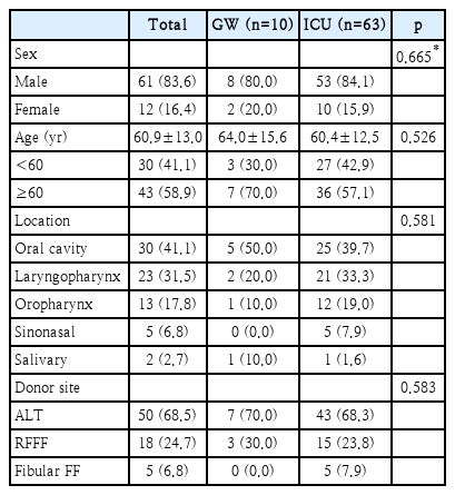
Clinical characteristics of both patient groups (ICU and GW protocol patients)
The total operation time and duration of microvascular anastomosis were shorter in the GW cohort (299.6±79.8 min and 64.4±11.5 min, respectively) than in the ICU cohort (478.9±126.5 min and 79.6±63.2 min, respectively). The difference of operation time was significant but not in microscopic time (p<0.001 and p=0.202, respectively). In the GW cohort, there was a tendency for larger flap size compared with that in the ICU cohort (11.6 cm and 10.2 cm, respectively) on average, although this difference was not significant (p=0.142) (Fig. 1).
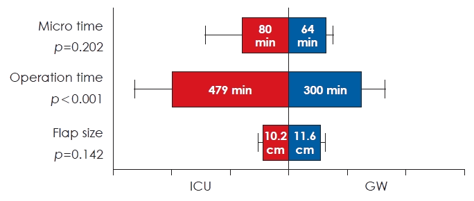
Intraoperative features comparisons between the two patient groups. Text in the graphs indicates the mean value in each group. The p-value is calculated by t-test. ICU, intensive care unit; GW, general ward.
ICU patients had significantly longer hospital stays compared with those of GW patients (25.9±24.2 days vs. 16.5±9.0 days; p=0.032). Days until resuming oral feeding and drain removal were shorter in the GW cohort (10.8 days and 8.2 days on average, respectively) than that in the ICU cohort (14.5 days and 8.7 days on average, respectively). However, the differences in days until oral feeding and drain removal between the two groups were not significant (p=0.225 and p=0.753, respectively) (Fig. 2).
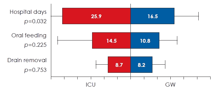
Recovery time (in days) between the two patient groups. The p-value for each test in the graphs stands for the p-value from the mean difference of each group using Student’s t-test. ICU, intensive care unit; GW, general ward.
All four indicators showed significant changes in accordance with the passage of postoperative time (p≤0.001 in “time” source of each) (Fig. 3 and Table 2).
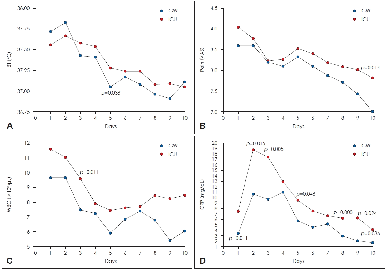
Hospital courses of postoperative day 1 to day 10 between ICU and GW groups. In each date, the p-value is displayed above the column when there is a significant difference in means in the t-test. Y-axis of each graph stands for (A) BT (°C), (B) pain in VAS, (C) WBC count (×103/μL), and (D) CRP (mg/dL) level. ICU, intensive care unit; GW, general ward; BT, body temperatur; VAS, visual analog scale; WBC, white blood cell; CRP, C-reactive protein.
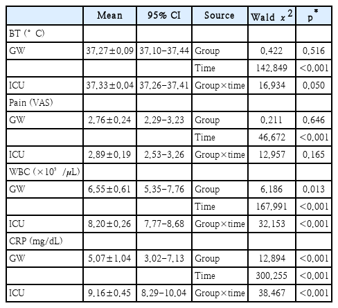
Significance test for each factor of hospital courses with GEE
Body temperature in the GW group was 37.27°C±0.09°C on average throughout the day, which was slightly lower than that observed in the ICU group (37.33°C±0.04°C) but was not significantly different (p=0.516 in “group” source) (Fig. 3A). However, the pattern of changes over time between the two groups differed significantly (p=0.050 in “group×time” source) (Fig. 3A and Table 2).
Pain (in VAS scale) in the GW group was 2.76±0.24 on average throughout the day, which was a slightly lower tendency than the pain (2.89±0.19) observed in the ICU group. This difference was not significant even when considering the patterns of changes over time (p=0.646 in “group” source and p=0.165 in “group×time” source, respectively) (Fig. 3B and Table 2).
The WBC count in the GW group was 6.55±0.61 (×103/μL) on average throughout the day, which was significantly lower that of 8.20±0.26 observed in the ICU group even when considering the patterns of changes over time (p=0.013 in “group” source and p<0.001 in “group×time” source, respectively) (Fig. 3C and Table 2).
The CRP level in the GW group was 5.07±1.04 (mg/dL) on average throughout the day, which was significantly lower than that of 9.16±0.45 observed in the ICU group even when considering the patterns of changes over time (p<0.001 in both “group” source and “group×time” source) (Fig. 3D and Table 2).
No significant difference was observed in complete flap failure rate when comparing ICU to GW protocol patients (3 of 63, 4.76% vs. 0 of 10, 0%; p>0.999). Furthermore, no significant difference was observed in the fistula formation rate between the ICU and GW patients (6 of 63, 9.52% vs. 1 of 10, 10%; p>0.999).
Given that operation time was the most prominent baseline difference between the two groups and a plausible confounder of postoperative recovery, we performed additional GEE analyses with adjustment for operation time. After adjustment, no significant group effect was observed for body temperature, pain, or WBC, whereas the group effect remained significant for CRP (Wald χ2=7.932, p=0.005). Similarly, the group-by-time interaction was no longer significant for body temperature or pain, but remained significant for WBC (Wald χ2=23.493, p=0.005) and CRP (Wald χ2=25.066, p=0.003). Operation time itself showed an independent association only with CRP (Wald χ2=6.862, p=0.009) (Supplementary Table 1).
Discussion
No significant differences were observed in flap failure or fistula formation between the ICU and GW groups. The GW group had a shorter operative time and shorter hospital stay, and unadjusted longitudinal analyses showed more favorable WBC and CRP trends in the GW group. Because operative duration was the most prominent between-group difference and a clinically relevant potential confounder, additional GEE analyses were performed with adjustment for operation time. After adjustment, the differences in body temperature and pain were attenuated, whereas more favorable longitudinal WBC and CRP trends persisted in the GW group. These findings suggest that part of the observed between-group differences was related to operative burden, but that GW-based postoperative management was at least not clearly worse than routine ICU care in selected patients, particularly with respect to flap-related outcomes.
Various explanations for the difference of postoperative course are provided in the literature. Mashrah, et al. [1] performed a systematic review on ICU vs. non-ICU care after head and neck free flap surgery of studies up to April 2021 and demonstrated that the incidence of postoperative pneumonia (p=0.018) and sepsis (p=0.033) were increased in the ICU setting. Indeed, hospital-acquired pneumonia is the most common infection in the ICU [6]. Ventilator-associated pneumonia is particularly associated with Pseudomonas aeruginosa infection in ICUs worldwide [7]. This frequently results from aspiration of oropharyngeal secretions containing colonized oral bacteria into the lungs [8]. Since head and neck flap surgeries frequently include airway and oral intervention, ICU environments could have particularly sensitive effects on infection.
Postoperative inflammatory markers, particularly CRP, may also reflect the degree of surgical trauma and perioperative stress, and the significantly longer operative time in the ICU group may have contributed to these differences [9]. In addition, differences in postoperative recovery pathways, including delayed extubation, overnight sedation with mechanical ventilation, and later mobilization, may also have affected pain and inflammatory trends [10,11]. ICU-related discomfort, frequent nursing interventions, and sleep disruption could likewise have influenced subjective pain scores. Therefore, these findings should be interpreted as multifactorial rather than as evidence of nosocomial infection alone.
Potential disadvantages of routine ICU care include increased cancellation of operations because of a lack of beds in the ICU and increased costs [12,13]. South Korea was ranked third lowest in terms of the number of ICU beds per population among high-income countries in 2017 [14]. The patient-to-nurse ratio in South Korea also has been reported to be as poor as 14.31 patients per nurse [5]. As South Korea is one of the most rapidly aging countries globally, the shortage of medical resources for patients with head and neck cancer will inevitably worsen. Therefore, we sought to establish a foundation for cost and time efficient GW (not even special ward) postoperative care for free flap head and neck surgery.
We acknowledge some limitations of our study, including bias due to historical ICU control and that the GW cohort had relatively few patients and was biased towards a specific period. This was mainly due to the ICU bed availability issues during the COVID outbreak period, when most patients were referred to the GW. Although no intentional selection was made based on clinical characteristics such as surgical difficulty, selection bias cannot be excluded in this retrospective study. Therefore, it would be inappropriate to conclude that GW care is safe for all free flap surgery patients. Instead, our findings suggest that GW-based postoperative management may be a feasible alternative to routine ICU care in selected patients, particularly those with a relatively uneventful intraoperative and immediate postoperative course, successful extubation, and shorter operative duration. In our cohort, operation time appeared to be both an important confounder and a clinically relevant factor associated with postoperative recovery. ICU care may still be more appropriate for patients with prolonged or complex operations, concern for airway compromise, or a need for closer immediate postoperative monitoring. Further studies with larger sample sizes are needed to validate these considerations and to explore interactions with other clinicopathological variables.
Supplementary Materials
The Supplement is available with this article at https://doi.org/10.3342/kjorl-hns.2026.00122.
Notes
Acknowledgments
AI-assisted technology (ChatGPT, OpenAI) was used only for language editing and clarity. All authors reviewed and edited the content and take full responsibility for the accuracy, integrity, and originality of the manuscript.
Author Contribution
Conceptualization: Doh Young Lee. Data curation: Hae Chan Park, Doh Young Lee. Formal analysis: Hae Chan Park. Investigation: Hae Chan Park. Methodology: Doh Young Lee, Hae Chan Park. Project administration: Doh Young Lee. Resources: Doh Young Lee. Supervision: Doh Young Lee. Validation: Doh Young Lee. Visualization: Hae Chan Park. Writing—original draft: Hae Chan Park. Writing—review & editing: Doh Young Lee.