A Case of Lateral Neck Desmoid-Type Fibromatosis Misidentified as Malignancy
악성 종양으로 오인된 경부 외측부 데스모이드형 섬유종증 1예
Article information

Abstract
Desmoid-type fibromatosis (DTF) is a rare, locally aggressive fibroblastic neoplasm with a high recurrence rate. We report a rare case of lateral neck DTF in a 37-year-old male. Imaging demonstrated a 7.7-cm mass infiltrating the sternocleidomastoid muscle with lymphadenopathy, mimicking malignancy. Due to an inconclusive biopsy, a wide excision with selective neck dissection was performed. Histopathology confirmed DTF with microscopic positive margins (R1) and nuclear beta-catenin positivity. Nine resected lymph nodes were negative of metastasis. Although initial postoperative imaging was clear, a 6-month follow-up CT identified a suspicious recurrence. Fine-needle aspiration showed no malignancy, and active surveillance was adopted after multidisciplinary discussion. This case highlights the diagnostic challenges of DTF mimicking lateral neck malignancy. It further supports the rationale for concurrent neck dissection when malignancy is suspected and the role of active surveillance following R1 resection in anatomically complex regions.
Introduction
Desmoid tumor, or desmoid-type fibromatosis (DTF), is a fibroblastic tumor arising from connective tissues such as muscle, fascia, and aponeuroses. Although it is classified histologically as a benign tumor because it lacks cellular atypia and metastatic potential, it shows marked local invasiveness into adjacent tissues and a high local recurrence rate of approximately 20%-39% after surgery [1]. Owing to these characteristics, it is classified as an “intermediate, locally aggressive” tumor in the World Health Organization classification of soft-tissue tumors [1].
The annual incidence has been reported to be 2-4 cases per million population worldwide, and DTF accounts for approximately 0.03% of all tumors [1]. DTF occurs predominantly in women in their 30s and 40s. DTF is classified by anatomic site as intra-abdominal, abdominal wall, or extra-abdominal. Extra-abdominal desmoid tumors account for approximately one-third of all cases. Head and neck involvement is relatively rare, accounting for approximately 7%-15% of cases, and the neck is the most commonly reported site within the head and neck region [2]. Approximately 80% of cervical desmoid tumors arise in the anterolateral neck [2]. Occurrence in the posterolateral or purely lateral neck, as in the present case, is very rare [2-4]. A Korean case of cervical DTF that was mistaken preoperatively for a malignant tumor and treated with concomitant neck dissection has been reported [5]. However, no Korean case of lateral neck DTF managed with active surveillance after R1 resection has yet been reported.
Because the head and neck region contains densely packed major neural and vascular structures, including the carotid artery, internal jugular vein, vagus nerve, and accessory nerve, it is difficult to balance complete resection with negative margins (R0) against functional preservation. In addition, preoperative imaging findings often resemble those of malignant soft-tissue tumors, making differential diagnosis challenging, and biopsy specimens alone frequently fail to yield a definitive diagnosis. We encountered DTF arising in the left lateral neck of a 37-year-old male. Preoperative CT demonstrated a large soft-tissue mass with cervical lymphadenopathy, raising suspicion of malignancy. Wide excision and selective neck dissection were performed, but the procedure resulted in R1 resection, and the subsequent clinical course raised suspicion of recurrence. We report this case together with a literature review of radiologic differential diagnosis, immunohistochemical diagnosis, the rationale for selective neck dissection, treatment strategies after R1 resection, and current guidelines.
Case
A 37-year-old male presented to the outpatient clinic with a mass below the left ear that he had noticed 1 year earlier. He had no significant pain or functional impairment but sought care because the mass appeared to have recently increased in size. His medical and family histories were unremarkable, and he had no history of trauma or neck surgery. On physical examination, a firm, non-tender, fixed mass measuring approximately 7–8 cm was palpated below the left ear. There were no overlying skin changes or other notable findings.
To evaluate the lesion in detail, contrast-enhanced neck CT and MRI were performed. CT revealed a 7.7-cm soft-tissue mass posterior to the left parotid gland, with invasion of the deep portion of the sternocleidomastoid muscle (SCM) and cervical lymphadenopathy, raising concern for malignancy (Fig. 1A and B). MRI showed an ovoid mass measuring 7.7×7.3×5.0 cm between the subcutaneous fascia and paraspinal muscles. On T2-weighted images, the mass showed heterogeneous high signal intensity reflecting a mixture of cellular and collagenous components (Fig. 1D). Contrast-enhanced T1-weighted images showed marked enhancement and infiltrative extension into the adjacent SCM (Fig. 1C).
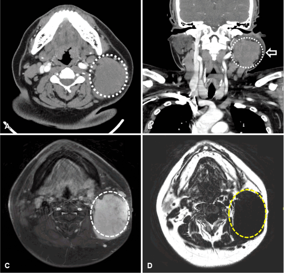
Preoperative imaging findings. Axial contrast-enhanced CT image shows a 7.7-cm heterogeneous soft-tissue mass in the left neck level II area (outlined by the dashed line), infiltrating the sternocleidomastoid muscle with associated regional lymphadenopathy (A). Coronal contrast-enhanced CT image demonstrates the cranio-caudal extent of the mass, outlined by the dashed line. The lesion abuts the sternocleidomastoid muscle (arrow) with an ill-defined interface, raising suspicion for an infiltrative soft-tissue neoplasm (B). Axial contrast-enhanced T1-weighted MR image demonstrates heterogeneous enhancement of the mass (outlined by the dashed line) (C). Axial fat-suppressed T2-weighted MRI image demonstrates heterogeneous high signal intensity and the extent of the lesion (outlined by the dashed line) (D).
Ultrasound-guided core needle biopsy was performed; however, the pathologic findings required differentiation from inflammatory myofibroblastic tumor (IMT) and low-grade fibromyxoid sarcoma (LGFMS), precluding a definitive diagnosis. Because a malignant soft-tissue sarcoma could not be excluded histologically and the enlarged lymph nodes could not be definitively characterized on imaging, nodal metastasis could not be ruled out. Intraoperatively, a left level II lymph node was the most suspicious based on its size and firmness. Given the possibility of nodal metastasis from sarcoma, selective neck dissection confined to left level II was performed together with wide excision of the left neck mass. The neck dissection was limited to level II to preserve uninvolved neck structures and minimize postoperative morbidity, while wide excision was performed in an attempt to achieve adequate oncologic clearance.
Final histopathologic examination identified a 12×9.5 cm DTF as the main lesion, with a satellite nodule (Fig. 2A). Histologically, relatively uniform spindle cells showed fascicular proliferation, with invasion of adjacent skeletal muscle and perivascular lymphocytic infiltration (Fig. 2B). Multifocal margin involvement was identified, with the largest focus measuring 0.5 cm; this corresponded to R1 resection (microscopically positive margins). In addition, a 0.8×0.8 cm DTF was identified in the mastoid tip-SCM ligament resection specimen, with margin involvement at that site as well. In the level II neck dissection specimen, all nine lymph nodes showed only chronic nonspecific lymphadenitis, with no evidence of metastasis.
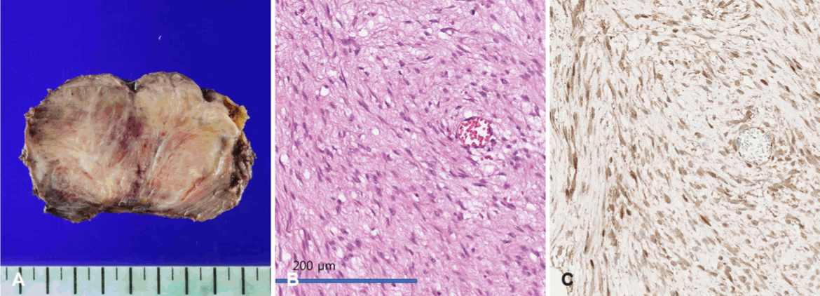
Histopathological and immunohistochemical findings. A: Gross specimen of the resected mass (12×9.5 cm), with a satellite nodule identified in the surgical specimen. B: Hematoxylin and eosin staining showing uniform spindle cell proliferation in fascicular pattern with skeletal muscle invasion (×200). C: Nuclear beta-catenin positivity on immunohistochemistry (×200). SMA positivity and S-100 negativity were also observed.
Immunohistochemistry showed nuclear beta-catenin positivity and SMA positivity, whereas S-100 was negative (Fig. 2C). The Ki-67 proliferation index was 4.49%, indicating low proliferative activity. These findings were consistent with sporadic DTF associated with a CTNNB1 gene mutation.
Neck MRI performed 3 months after surgery showed only postoperative changes at the surgical site, with no evidence of residual tumor. However, neck CT performed 6 months after surgery revealed a new irregular enhancing lesion measuring 1.1×1.9 cm at left level II, raising the possibility of residual or recurrent tumor (Fig. 3). Fine-needle aspiration (FNA) was therefore performed, and cytology showed clusters of benign spindle cells. The cytologic findings could represent either fibromatosis or reactive stromal cells, precluding a definitive diagnosis. No malignant cells were identified.
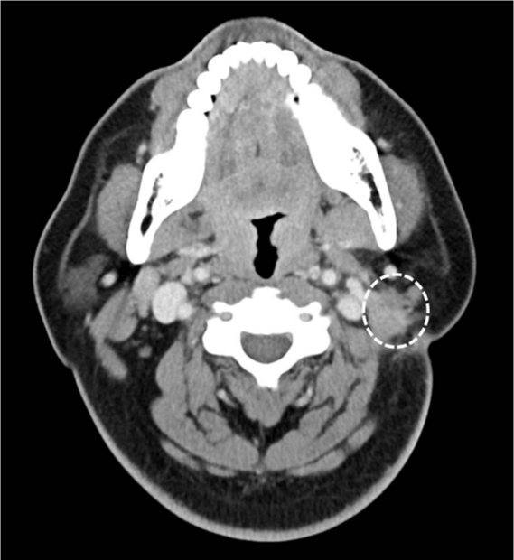
Contrast-enhanced neck CT performed 6 months postoperatively. Axial contrast-enhanced CT image demonstrates a newly developed 1.1×1.9 cm irregular enhancing lesion at the surgical bed (outlined by the dashed line). Because the lesion could not be definitively differentiated from postoperative fibrosis or residual/recurrent desmoid-type fibromatosis, the patient was managed with continued active surveillance.
Following multidisciplinary discussion, active surveillance was selected rather than immediate additional treatment, and follow-up neck CT was planned after 3 months.
This study was exempt from review by the Institutional Review Board (IRB) of Gyeongsang National University Changwon Hospital (IRB No. GNUCH 2026-04-017), and the requirement for patient consent was also waived.
Discussion
In soft-tissue tumors of the head and neck, the low diagnostic yield of biopsy is attributable not only to insufficient specimen volume but also to morphologic overlap among various spindle-cell tumors [1]. When a definitive diagnosis cannot be reached on preoperative core needle biopsy, as in this case, differentiation from IMT, LGFMS, and high-grade fibrosarcoma is essential.
On imaging, high-grade fibrosarcoma is frequently accompanied by aggressive infiltration, extensive central necrosis, and bone destruction, whereas DTF rarely exhibits internal necrosis or bone destruction despite its marked infiltrative nature [6]. In addition, IMT tends to be relatively well circumscribed and to show homogeneous enhancement, whereas DTF has indistinct borders with adjacent muscle and demonstrates characteristic heterogeneous signal intensity on T2-weighted images according to the proportions of cellular and collagenous components [6]. Definitive diagnosis relies on immunohistochemical analysis: IMT can be differentiated by ALK expression [7], and LGFMS shows strong positivity on MUC4 immunostaining [8]. In contrast, DTF is histologically distinguished by fascicular proliferation of uniform spindle cells without cellular atypia in a collagenous stroma, nuclear beta-catenin positivity, and SMA positivity [1].
The management of DTF after R1 resection has changed substantially in recent years. Previously reported Korean and international cases of lateral and posterolateral neck DTF frequently underwent R1 resection because of anatomic complexity. Previous reports have described recurrence during observation alone [3] and the use of adjuvant radiotherapy or delayed radiotherapy after progression to reduce local recurrence (Table 1) [2,4]. In the present case, the 7.7-cm mass infiltrated adjacent critical neurovascular structures, and complete resection was limited by the need to preserve function. R1 resection was therefore unavoidable. In the present case, active surveillance was adopted in accordance with current guidelines following R1 resection, without initiating immediate adjuvant treatment, representing a clear point of distinction from previously reported Korean cases. The patient has remained clinically stable [5].
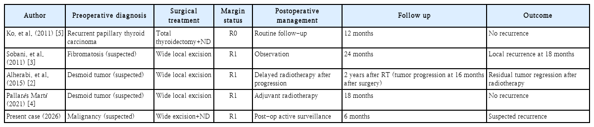
Clinical characteristics and outcomes of lateral neck desmoid-type fibromatosis cases in literature
Recent guidelines, including those of the European Society for Medical Oncology (ESMO) and the global consensus, strongly recommend “primary active surveillance,” in which surgery itself is deferred, as the first-line approach for asymptomatic patients [9]. In a study using a front-line conservative approach, Fiore, et al. [10] reported that a substantial proportion of patients achieved long-term stabilization or spontaneous regression without surgery, and a prospective observational study by Colombo, et al. [11] likewise found spontaneous regression in some patients during active surveillance, supporting conservative treatment strategies based on individual risk and anatomic characteristics rather than initial aggressive surgery. The practical clinical significance of this case lies in demonstrating that, even when initial surgery and R1 resection were unavoidable due to preoperative suspicion of malignancy, transitioning to “postoperative active surveillance” once final pathology confirms DTF is a reasonable and justifiable approach, rather than pursuing extensive re-resection or adjuvant radiotherapy. This approach is consistent with current guidelines favoring conservative management to reduce unnecessary functional morbidity [3,11]. Although CT detected a lesion suspicious for recurrence 6 months after surgery, active surveillance was continued in view of the limitations of FNA in distinguishing fibromatosis from postoperative reactive stromal cells and the unpredictable natural history of DTF.11) If definite tumor growth is identified on future follow-up imaging, biopsy will be repeated and a multidisciplinary treatment plan, including re-resection, radiotherapy, or systemic therapy such as sorafenib, will be reconsidered [12].
In conclusion, large DTF arising in the lateral neck is difficult to diagnose definitively before surgery. When lymphadenopathy is present, wide excision and neck dissection comparable to those used for malignancy may be undertaken before a definitive diagnosis is established. However, if the diagnosis is established preoperatively, neck dissection can be avoided because of the nonmetastatic nature of the disease. When R1 resection is confirmed after surgery or recurrence is suspected, active surveillance should be prioritized in accordance with current clinical guidelines. Follow-up findings and the limitations of FNA should be considered together when developing an individualized multidisciplinary treatment plan.
Notes
AI-assisted Translation
Generative AI was used for English translation. The final version was reviewed and approved by the authors and the Editorial Office, and the authors retain final responsibility for the content.
Bilingual Publication
This article is available in both Korean and English.
Acknowledgments
None
Author Contribution
Conceptualization: Seulki Song. Data curation: Seulki Song. Formal analysis: Jae Hoon Kim. Investigation: Jae Hoon Kim. Methodology: Jae Hoon Kim. Project administration: Jin Pyeong Kim, Seulki Song. Resources: Jae Hoon Kim. Software: Seulki Song. Supervision: Seulki Song, Jin Pyeong Kim. Validation: Jin Pyeong Kim, Dae Hyun Song, Seulki Song. Visualization: Jae Hoon Kim. Writing—original draft: Jae Hoon Kim. Writing—review & editing: all authors.