성인 환자에서 발생한 비익의 점액종
Myxoma of Nasal Ala in an Adult Patient
Article information

Trans Abstract
Myxoma most commonly occurs in the atria, but is rare in the nasal cavity. A 58-year-old male patient presented with a nasal mass. We used endoscopic endonasal approach for treatment, and the patient was finally diagnosed with nasal ala myxoma. We report here a rare case of a patient with nasal ala myxoma along with a literature review.
Introduction
Myxomas are benign neoplasms that were first reported by Virchow in 1871 [1,2]. Myxomas most frequently occur in the atria, but can also be found in bone, skin, skeletal muscle and subcutaneous tissue [2-5]. Myxomas of head and neck are rare and occur in the mandible, maxilla and soft tissue of the face [2,5]. Herein, we reported an unusual case of a patient with myxoma at the right nasal ala which was resected using endoscopic endonasal approach.
Case
A 58-year-old male patient visited our hospital with a right nasal dorsum swelling. CT scan demonstrated a 1.3×0.5 cm poorly enhanced low attenuation lesion in right nasal ala (Fig. 1). Fine-needle aspiration cytology tests confirmed benign myxoid spindle cell lesions. Surgery was recommended, but the patient refused the operation. Five years later, the patient returned to the hospital because the swelling of the right nose worsened (Fig. 2). MRI demonstrated a 1.7×1.3×1.2 cm T1-weighted image low signal intensity, T2-weighted image high signal intensity, and peripheral enhanced soft tissue lesion in right nasal ala (Fig. 3).
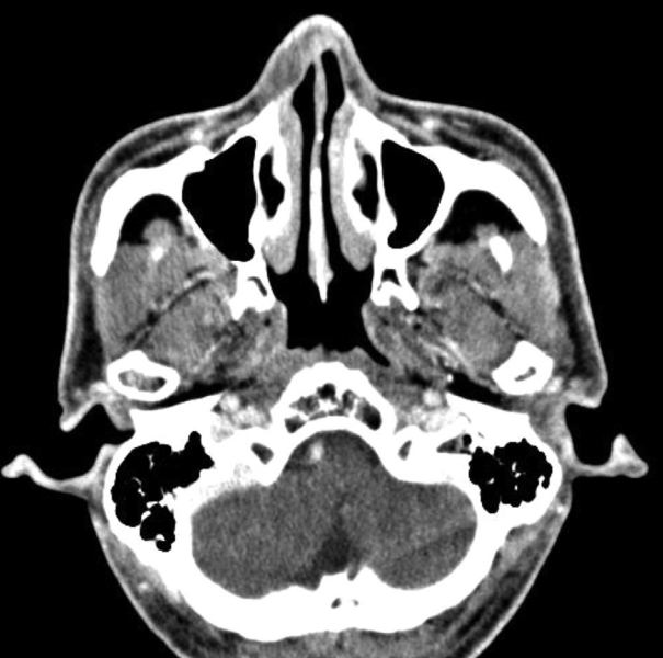
A 1.3×0.5 cm poorly enhanced low attenuation lesion in right nasal ala.
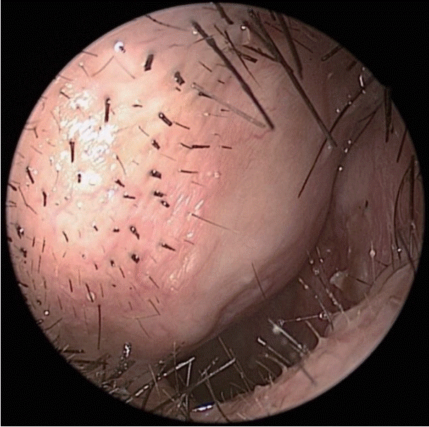
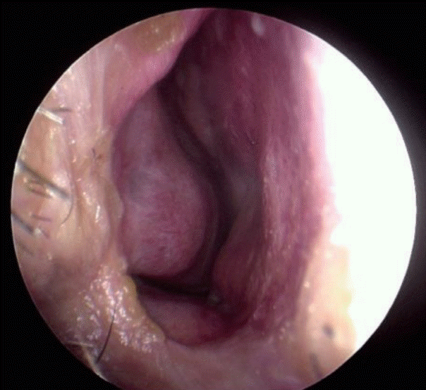
Nasal endoscopy showing a swelling lesion in right nasal alar.
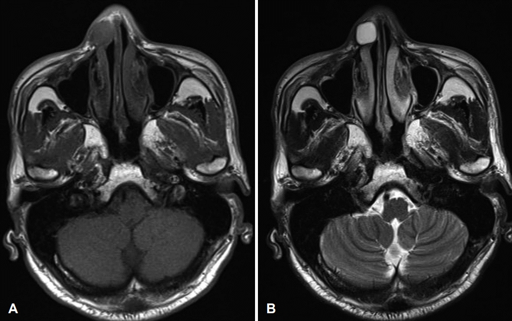
A 1.7×1.3×1.2 cm T1-weighted image (A) low signal intensity, T2-weighted image (B) high signal intensity, peripheral enhanced soft tissue lesion in right nasal ala.
We performed endoscopic mass removal through the right nasal cavity. After an incision was placed in the right ala, the mass and the mucous membrane surrounding the mass were carefully peeled off and completely removed (Fig. 4). Histological examination confirmed the diagnosis of a myxoma (Fig. 5). After 9 months of surgery, patient is being followed up without complications or cosmetic problems (Fig. 6).
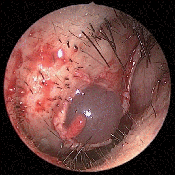
Intraoperative photo showing a myxoma in right nasal alar.
Slide reveals hypocellular myxoid stroma with scant blood vessels. A few spindle cells show bland ovoid nuclei and no mitotic activity (hematoxylin and eosin stain, ×100).
There are no abnormal findings in the nasal endoscopy performed 9 months after surgery
Discussion
Myxoma of the head and neck is a very rare benign neoplasm [1-8]. Myxoma is most frequently found in the atria, but can also be found in bones, skeletal muscle, skin, and soft tissue [4,5]. Myxoma of the head and neck occurs in patients between the ages of 1 and 73 years, but most often occurs in the second to fourth decades [6]. In the head and neck, bony myxoma is more common than soft tissue myxoma [4]. Myxomas of the head and neck occur mainly in facial bones such as the mandible or maxilla [2]. Soft tissue myxomas of head and neck characteristically grow by local invasion and expansion and do not undergo metastasis [2-5].
The most common symptom in patients with myxoma is a mass that grows slowly over several years [4]. The characteristic clinical symptom of paranasal myxoma is painless swelling in the area, usually around the nasolabial or paranasal region [2,6]. Common symptoms include nasal congestion, nasal obstruction and nasal bleeding [8].
Radiographically, myxomas are usually well-defined ovoid masses with bony destruction or remodeling that show attenuation similar to that of water on CT [2]. On MRI, the tumor exhibits the signal intensity characteristic of fluid [6]. Intraoperatively, most masses are usually identified as glistering gelatinous masses which are oval or spherical in shape [2,4,7]. Histologic features of myxoma include spindled and stellate cells embedded in a fibromyxoid background [2,5,7,8]. Therefore, the definitive diagnosis of myxoma is made by microscopic examination to identify bland spindle cell myxoid lesion [2-6]. The spindled cells of myxoma will stain positively for vimentin and may show some positivity for S-100 protein and muscle-specific actin [2].
Surgical resection is the first treatment option for myxoma [1-8]. Inadequately defined tumor boundaries are associated with recurrence [4-6]. Conservative surgical techniques, such as, enucleation or curettage have a higher rate of local recurrence than more aggressive surgeries, such as, wide en-bloc resection [2,4,5]. However, aggressive surgery on the head and neck can be aesthetically undesirable as it leaves scars. The endoscopic endonasal surgery is a minimally invasive technique that leaves no external scars and can greatly reduce the possibility of deformity. At the same time, the endoscopic nasal approach can better identify tumor boundaries and secure sufficient surgical margins, thereby reducing the probability of recurrence.
In conclusion, although myxoma of the nasal cavity is very rare, it should be differentiated from other tumors occurring in the nasal cavity. Endoscopic endonasal surgery is considered a good treatment option for nasal ala myxoma for cosmetic reasons.
Acknowledgements
None
Notes
Author Contribution
Conceptualization: Dong Hoon Lee. Data curation: Bong-Jin Shin, Keon Woo Park, Dong Hoon Lee. Formal analysis: Dong Hoon Lee. Investigation: Dong Hoon Lee. Supervision: Sang Chul Lim. Validation: all authors. Writing-original draft: Bong-Jin Shin, Dong Hoon Lee. Writing-review & editing: all authors.