백반증 양상의 성문 아밀로이드증 1예
A Case of Glottic Amyloidosis Presenting as Leukoplakia
Article information
Trans Abstract
Amyloidosis is defined as a deposit of amyloid substance. While it rarely occurs in the head and neck region, it is most commonly found in the larynx. Laryngeal amyloidosis can occur in the false vocal cord, ventricle, and glottis etc. The typical feature of laryngeal amyloidosis is a round yellowish submucosal mass. A 72-year-old male presented with voice change that began a couple of years ago. The rigid laryngoscopy showed a whitish patch in the medial and superior surfaces of the left true vocal fold. He was pathologically diagnosed with amyloidosis by laryngeal microsurgery. With a relevant review of literature, we report this case as it demonstrates rare, atypical features of laryngeal amyloidosis.
서 론
아밀로이드증(amyloidosis)은 “아밀로이드”라는 부정형 단백질이 세포 외 공간에 비정상적으로 침착되어 발생하는 질환이다[1]. 원인 미상인 일차성, 다른 질환과 연관되어 발생하는 이차성 등으로 분류할 수 있다[2]. 두경부 아밀로이드증은 주로 일차성으로 후두에 호발하며, 구강설, 갑상선, 인두 등에도 발생할 수 있다[3,4]. 후두에서는 가성대, 후두실, 성문 및 성문 하부 등에 발생할 수 있으며, 전형적인 임상 소견은 매끈한 황색의 점막 하 종물이다[3-5].
72세 남자가 내원 2년 전부터 시작된 음성 변화를 주소로 내원하였다. 후두내시경에서 좌측 진성대에 백반증이 관찰되어, 후두미세수술을 시행하여 일차성 아밀로이드증으로 최종 진단되었다. 저자들은 매우 드문 백반증 양상의 성문 아밀로이드증을 경험해서 문헌 고찰과 함께 보고하고자 한다.
증 례
72세 남자 환자가 내원 2년 전부터 시작되고, 최근 1개월 동안 심해진 음성 변화를 주소로 일차 의료기관에서 의뢰되었다. 고지혈증, 고혈압 및 당뇨 등의 질환이 있고, 흡연력은 10갑년으로 20년 전부터 금연한 상태이며, 음주력은 미미하였다. 객담, 기침 및 인후두 역류 증상 등은 없었다. 경성 후두 내시경 검사에서 우측 진성대에 성대구 및 좌측 진성대의 내측면과 상연 등에 백색반이 관찰되었다(Fig. 1A). 성대진동검사에서 점막 파동은 양측 모두 감소되어 있었고, 성대 내전 시 성문 틈이 관찰되었다. GRBAS 척도 평정법에서는 대부분 3점이었고, 최장 발성 지속시간은 5.3초로 감소된 양상이었다. 다면음성검사(Multi-Dimensional Voice Program, PENTAX Medical, Montvale, NJ, USA)에서 기본 주파수가 147.68 Hz, 주파수 변동률(jitter)은 1.75% (참고치<1.1%), 진폭 변동률(shimmer)은 7.41% (참고치<3.8%), 잡음 대 배음비(noise to harmonic ratio)는 0.164 (참고치<0.2) 등으로 잡음대 배음비를 제외한 주요 평가 지표가 증가된 소견이었다.

The rigid laryngoscopic findings. A: It shows deep sulcus on right true vocal fold (arrows), leukoplakia was observed on the medial and superior surfaces in left true vocal fold (arrowheads). B: At the postoperative one day, there was no bleeding in operation site. C: At the postoperative 4 months, it shows well healed lesion and no evidence of leukoplakia.
환자가 고령이고, 내원 3개월 전에 작성된 일차 의료기관의 의뢰서에서도 동일한 부위에 백반증이 기술된 점 등을 고려하여, 백반증의 호전 가능성이 적다고 판단되어 후두미세수술을 계획하였다. 환자에게는 술전에 우측 진성대에 성대구증, 좌측 진성대에 백반증 및 노령에 의한 발성장애가 있어서, 술후 음성 호전이 어려울 가능성이 높다고 설명했다. 우측 성대구증은 수술 없이 경과 관찰만 하겠으며, 좌측 백반증은 술후 음성이 악화되는 경우도 흔하다고 설명했다.
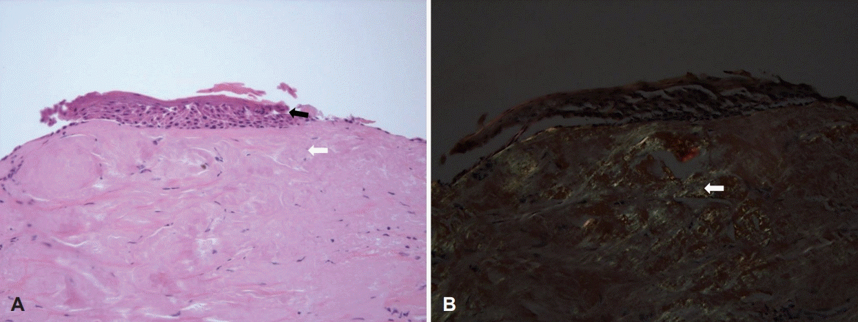
수술은 현미경 시야에서 좌측 진성대의 내측면과 상연 등에 백색반이 관찰되어, 미세수술용 겸자와 가위 등을 이용한 점막 박리법(mucosal stripping)으로 병변을 제거하고, 에피네프린 거즈를 위치시켜 지혈하고 수술을 종료하였다. 술후 1일에 후두내시경 소견에서 수술 부위의 이상 소견은 없었고, 환자는 음성안정 및 주의 사항 교육 후 퇴원하였다(Fig. 1B). 조직병리 소견은 hematoxylin and eosin (H&E) 염색 200배에서 점막의 과각화증 소견과 점막 하부에 부정형의 균일한 호산성 물질이 다량으로 침착된 양상이었고(Fig. 2A), 편광 현미경으로 관찰한 Congo-red 염색 200배에서 밝은 황록색의 이중 굴절 소견이 관찰되어 아밀로이드증으로 확진되었다(Fig. 2B). 국소성과 전신성 아밀로이드증을 감별하기 위하여 류마티스 내과로 의뢰되었고, 선행된 검사와 중복되지 않는 혈액 검사, 뇨단백 전기영동검사, 결핵반응검사, 심장 및 복부 초음파 등을 시행하였으며, 전신성을 의심할 수 있는 소견이 없어서 일차성 국소형 아밀로이드증으로 최종 진단되었다. 술후 2주째 후두내시경 검사에서 수술 부위 점막의 정상 치유 과정이 관찰되고, 술후 4개월에 시행한 검사에서 병변의 재발 소견은 없었다(Fig. 1C). 음성 안정을 하면서 술후 1개월에 1주 간격으로 3회의 음성 치료를 시행하였지만, 술후 8주에 시행한 음성검사에서는 주요 지표에서 술전과 유사한 결과를 보였고, 10개월이 경과한 현재까지 재발 소견 없이 추적 관찰 중이다.

The microscopic findings of specimen. A: Hyperkeratotic squamous epithelium (black arrow), subepithelial amyloid deposits and subepithelial pinkish amorphous deposits were observed (white arrow) (hematoxylin and eosin, ×200). B: Amyloid deposits showed apple-green colored birefringence in polarized microscopy (white arrow) (Congo-red stain, ×200).
고 찰
아밀로이드증은 기저막 주변의 세포 사이 간격에 불용성 부정형 단백질인 아밀로이드가 침착되어, 조직에 영향을 주어 장기의 기능 저하를 유발하게 된다[6]. 아밀로이드는 격자 모양의 베타판(β-pleated sheet) 형태의 섬유소로 이루어졌고, 이중 95%가 원섬유 단백질이고, 나머지는 알파당 단백(α-glycoprotein)으로 구성되어 있다[7]. 임상적으로는 일차성과 이차성 아밀로이드증, 국소성과 전신성인 골수 관련 및 유전 관련 아밀로이드증 등으로 분류할 수 있다[2].
두경부 아밀로이드증은 100만 명당 1-2명에서 발생하는 드문 질환으로 대부분 일차성이며, 후두, 구강설, 갑상선, 인두 등에 발생한다[4]. 후두에서는 모든 부위에 발생할 수 있으며, 성문상부 및 성문부 등이 하부보다 비교적 흔하다[8,9]. 후두내시경 소견은 황색 상 결절이 가장 흔하지만, 다양한 색상으로 보일 수도 있으며, 진행된 병변에서는 붉은 용종 형태로 나타날 수 있다[9]. 또한 가성대 및 성문하부에서는 산재성, 성문에서는 국소적 양상으로 관찰된다[9]. 임상 증상은 발생 부위에 따라 비특이적인 증상을 보이며, 대부분 병리 조직검사로 확진된다[10].
본 증례의 주 증상인 음성 변화는 후두미세수술 전후로 비슷한 것으로 보아서, 애성은 우측 성대구증이 주 원인으로 생각된다. 조직병리 소견은 H&E 염색에서 다량의 부정형 호산구성 단백질이 세포 간격에 축적을 보이고, Congo-red 염색에서 편광현미경으로 밝은 황록색의 이중 굴절 소견이 특징적으로 나타난다[7]. 후두 아밀로이드증으로 진단되면 반드시 이차성 및 전신성 아밀로이드증에 대한 감별을 위해 혈액검사, 혈액 및 소변 단백의 면역전기영동검사, 심전도검사, 흉부 및 두부 방사선학적 검사, 심장 및 복부초음파 등을 반드시 시행해야 하고, 필요한 경우 골수검사를 할 수 있다[1]. 본 증례와 같은 국소성 아밀로이드증은 수술로 병변이 완전히 제거되면 예후가 양호하지만, 재발의 가능성 때문에 추적 관찰이 필수적이다[7].
양측 가성대 및 진성대 등에 황색 결절을 보인 증례에서 제자리 암종을 동반한 아밀로이드증의 보고가 있었고, 백반증이 종종 제자리 암종으로 진단되는 것을 고려하면, 후두 백반증과 아밀로이드증의 연관성에 대한 연구도 필요해 보인다[5]. 그러나, 현재까지 아밀로이드증 자체가 암종으로 발전될 수 있다는 보고는 없다. 본 증례는 후두내시경과 수술 현미경 모두에서 일반적인 백반증의 소견이었는데, 저자들의 검색으로는 이러한 양상의 성문부 아밀로이드증은 보고가 없었던 것으로 생각된다[11]. 저자들은 본 증례를 통해서 임상적으로 흔히 경험하는 후두 백반증의 감별진단에 드물지만 아밀로이드증의 가능성도 염두에 두면서 술전 평가 및 수술 등을 진행해야 한다는 교훈을 얻었다.
Acknowledgements
None
Notes
Author Contribution
Conceptualization: Seung Woo Kim. Data curation: Seong Kyu Moon, Mi Ji Lee. Formal analysis: Moon Seung Beag. Investigation: Seong Kyu Moon. Methodology: Seung Woo Kim, Mi Ji Lee. Software: Seong Kyu Moon. Supervision: Seung Woo Kim. Validation: Moon Seung Beag. Visualization: Moon Seung Beag. Writing—original draft: Seong Kyu Moon, Mi Ji Lee. Writing—review & editing: Seung Woo Kim, Mi Ji Lee.
