하비갑개에 발생한 고도 B 세포 림프종
A Case of High-Grade B Cell Lymphoma, Not Otherwise Specified Originating From Interior Turbinate
Article information

Trans Abstract
High-grade B cell lymphoma not otherwise specified (HGBL, NOS) is a new diagnostic category of non-Hodgkin lymphoma (NHL) based on World Health Organization statement in 2016. Up to 40% of NHLs are located in the extra-nodal area, commonly involving the gastrointestinal tract, head and neck region, bone and skin. The diagnosis of NHL originating from the inferior turbinate (IT) is rare, and there have been no reported cases yet of HGBL, NOS in IT. In this study, we report on a 43-year-old female who had a nasal polyp in IT and was diagnosed with HGBL, NOS after surgery. Endoscopic polypectomy under general anesthesia was performed. A histopathological report confirmed the resected specimen as HGBL, NOS, with a clear resection margin. Nasal obstruction was improved immediately after surgery and the patient was followed up for 10 months without any recurrence or complications.
서 론
고도 B 세포 림프종(high-grade B cell lymphoma, HGBL)은 2016년에 개정된 세계보건기구(World Health Organization, WHO)의 분류 기준에 따라 MYC와 BCL2 또는 BCL6의 유전자 재배열을 가지는 고도 B 세포 림프종과 이외의 달리 분류되지 않은 고도 B 세포 림프종(HGBL, not otherwise specified; HGBL, NOS)으로 새롭게 분류되었다[1]. 기존의 비호지킨 림프종은 악성림프종의 60%를 차지하였고 두경부 영역에서 발생하는 악성종양의 1% 미만으로 유발되는 것이 보고되었다. 비호지킨 림프종의 대부분은 림프절에 국한되어 발생하지만 40%에서 림프절 외 침윤을 보인다[2,3]. 하지만 새롭게 정의된 달리 분류되지 않은 고도 B 세포 림프종은 매우 드물게 발생하고, 림프절 외 침윤을 보이는 증례는 아직까지 국내에서는 보고되지 않았다. 특히 하비갑개에 발생한 경우는 국내외에서 보고된 증례가 아직까지 없었다.
따라서 본 증례는 하비갑개에서 발생한 종물을 비용종으로 생각하고 수술을 시행하였으나 수술 후 달리 분류되지 않은 고도 B 세포 림프종으로 진단하고 치료한 경험을 보고하고자 한다.
증 례
43세 여자 환자가 내원 1개월 전부터 시작된 우측 코막힘과 우측 점액성 콧물을 주소로 본원 이비인후과에 내원하였다. 환자는 특이 과거력 및 가족력이 없었으며 전신증상(B symptom)은 호소하지 않았다. 비내시경 소견에서는 경계가 불규칙한 종물이 우측 하비도의 후방을 채우며 후비공까지 진행되어 있었고 우측으로 비중격만곡증 소견을 보였다(Fig. 1).
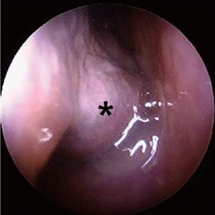
Preoperative endoscopic view of right nasal cavity. A polypoid mass (asterisk) fulling the inferior meatus is located between inferior turbinate and nasal septum.
수술을 계획하기 전에 해부학적 이상 유무를 확인하기 위해 부비동 전산화단층촬영(ostiomeatal unit CT)을 시행하였고, 우측 하비갑개의 후방에서 기원한 2.2 cm 크기의 폴립 양상의 종물이 관찰되었다. 이 종물은 후비공까지 커져 있었으나 하비갑개와 상악동 내측 벽의 골결손이나 그 이외 부비동 및 비강 내 특이소견은 관찰되지 않았다(Fig. 2).
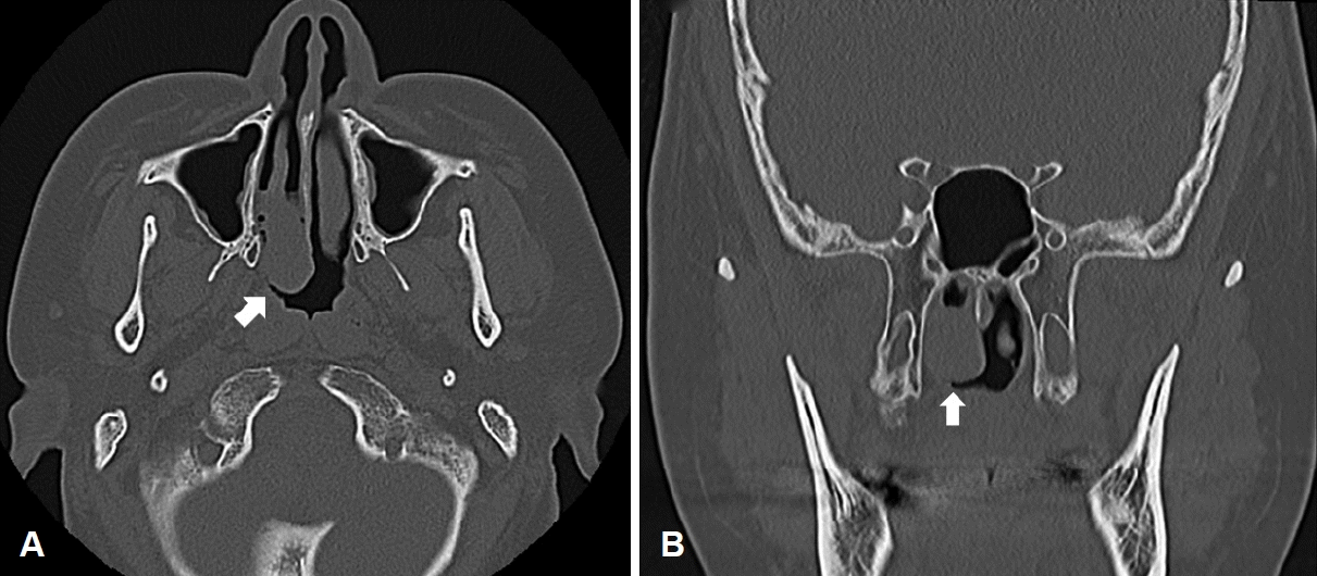
Preoperative ostiomeatal unit CT can. A: Non enhanced axial CT scan shows 2.2 cm sized polypoid lesion (arrow) in posterior aspect of right inferior turbinate, bulging to right posterior choana. B: Non enhanced coronal view of mass (arrow) within right nasal cavity shows no evidence of invasion into surrounding bony structures.
우측 하비갑개에서 기원한 단순한 비용종으로 생각하고 전신마취 후 비내시경을 이용하여 종물의 완전 절제를 계획하였다. 우선 비중격교정술 및 하비갑개 외향골절술을 시행하여 수술 시야를 확보하였고 종물은 하비갑개의 후하방 점막에서 기원하는 것을 확인하였다. Sickle knife와 Freer elevator 그리고 cutting forcep을 이용하여 종물이 부착된 주변 점막을 포함한 완전 절제를 시행하였고 2.2×2×1 cm 크기의 연조직 종물이 확인되어 조직검사를 진행하였다.
조직병리학적 검사 결과는 절제된 조직 내 모세포와 유사한 형태(blastoid)인 산재한 염색질과 뚜렷한 핵소체를 가지는 크거나 중간 크기의 림프구 세포 증식 소견과 함께 다수의 유사분열이 관찰되었다. 추가로 시행한 면역 조직 화학검사에서는 CD79a, CD20, CD5 Bcl-2, Bcl-6 양성을 보였다. 또한, CD23, CD21, CD4, CD3, cycllin D1은 음성을 보였으며 ki-67 labeling index가 90% 이상으로 확인되었다. 하지만 추가로 시행한 FISH 검사에서는 MYC와 BCL2, BCL6의 유전자 전위는 확인되지 않아 최종적으로 달리 분류되지 않은 고도 B 세포 림프종으로 진단되었다(Fig. 3).
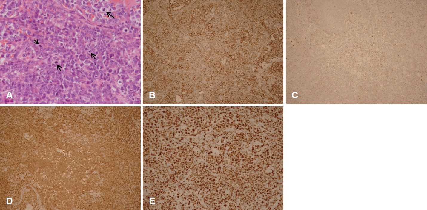
Histopathologic findings. A: Diffuse proliferation of large to medium sized atypical lymphoid cells with vesicular nuclei showing prominent nucleoli. Note frequent mitosis (arrow) (hematoxylin and eosin, ×200). B: Tumor cells are positive for CD20 (immunohistochemistry staining, ×100). C and D: Tumor cells are positive for Bcl-2 (immunohistochemistry staining, ×100) and Bcl-6 (immunohistochemistry staining, ×100). E: Ki-67 labeling index reveals about 90% (immunohistochemistry staining, ×100).
환자는 수술 후 다음 날 퇴원하였고 수술 전에 호소하던 우측 코막힘과 점액성 분비물 증상은 호전되었다. 이후 혈액종양내과로 전과되어 골수 검사를 진행하였고 골수로의 전이는 관찰되지 않았으며 양전자방출 컴퓨터단층촬영검사(PET-CT)에서 원격전이 소견은 확인되지 않아 앤아버 병기(Ann Arbor staging)상 stage I로 최종 진단하였다. 비강 내 잔존병변의 치료를 위해 총 3주기의 rituximab, cyclophosphamide, doxorubicin, vincrinstine, prednisone (R-CHOP) 항암치료와 총 방사선량 3000 cGy의 방사선 요법을 시행하였다. 수술 후 10개월간 외래에서 경과 관찰하였고 처음 내원 시 호소하였던 증상은 호전되었다. 또한, 비내시경에서 합병증과 재발 소견이 관찰되지 않았고 추가적으로 시행한 부비동 CT와 PET-CT에서도 재발 소견이 없어 정기적 추적 관찰 중이다(Fig. 4).

Follow-up endoscopic examination 10 months after surgery. The resection site is well healed with no sign of disease or complications.
고 찰
미분류 고도 B 세포 림프종은 2008년 WHO 분류에 따라 미만성 거대 B 세포 림프종과 버킷림프종의 중간적인 세포학적 특징을 가지고 임상적으로 악성도가 높은 성숙 B 세포 림프종이다[2]. 미분류 고도 B 세포 림프종의 다수는 MYC, BCL2 재배열을 가지는 이중유전자 이상을 보이거나 추가적으로 BCL6 재배열을 보이는 삼중유전자 이상을 나타냈는데 이런 경우에는 임상적으로 악성도가 높았으며 R-CHOP과 같은 기존의 항암치료에 반응이 좋지 않았다[4,5]. BCL2는 항세포자멸사 기능을 수행하는 단백질로, 과발현 될 경우 B 세포의 생존에 유리하게 작용한다고 알려져 있다[5]. 또한 BCL6는 p53과 밀접한 관계를 가지는 전사 억제 인자로, 과발현될 경우 DNA 손상에 대한 세포 자멸사를 막는다[5].
하지만 이중 혹은 삼중유전자 이상을 가지는 미분류 고도 B 세포 림프종은 미만성 거대 B 세포 림프종과 버킷림프종의 중간적인 특성으로 인해 정확한 진단과 치료에 어려움이 있는 경우가 발생하였다[6]. 이에 따라 2016년 WHO에서는 고도 B 세포 림프종에 대하여 새롭게 분류 개정하였고, 이는 MYC와 BCL2, 그리고/또는 BCL6의 재배열을 가지는 고도 B 세포 림프종(HGBL with MYC and BCL and/or BCL6 rearrangements)과 MYC와 BCL2, 그리고/또는 BCL6의 재배열을 가지지 않는 달리 분류되지 않은 고도 B 세포 림프종(HGBL, NOS)으로 분류하였다[1,7]. 비·부비동에 발생하는 B 세포 림프종은 드물게 발생하지만 과거 진단기준이 개정되기 전에 전 세계적으로 보고되어져 왔다. 대부분은 미만성 거대 B 세포 림프종이였으며 국내에서도 시력소실을 동반한 거대한 부비동 미만성 거대 B 세포 림프종 1예가 보고되었다[8]. 진단 기준이 새롭게 개정된 후에 달리 분류되지 않은 고도 B 세포 림프종은 진단 자체가 매우 드물어 아직까지 이 병에 대한 원인과 병리기전이 밝혀지지 않았다. 현재까지 국내에서는 보고된 증례가 없으며 국외에서는 Potts 등[9]이 보고한 대동맥 주위 림프절에 발생한 1예와 Brody 등[10]이 보고한 상악동에 발생한 1예가 있으나 하비갑개에 발생한 경우는 아직까지 전 세계적으로 보고된 적이 없었다.
림프종은 조직학적인 진단이 중요하지만, 고도 B 세포 림프종은 미만성 거대 B 세포 림프종, 미만성 거대 B 세포 림프종과 버킷림프종의 중간형태, 모세포와 유사한 형태(blastoid), 림프모구 림프종 등 다양한 조직학적 소견을 보일 수 있어 감별을 위하여 면역조직화학검사가 반드시 필요하다. 특히 달리 분류되지 않은 고도 B 세포 림프종은 조직학적으로 거대 B 세포 림프종과 버킷림프종의 중간 형태를 보이거나 모세포와 유사한 형태로 보인다[1]. 본 증례에서는 조직학적으로 모세포와 유사한 형태를 보였으며 이에 고도 B 세포 림프종과 B 세포 림프모구 림프종 등을 감별하기 위해 골수 조직검사 및 면역조직화학검사를 시행하였다[11,12]. 골수 조직검사에서 이상소견이 없어 B 세포 림프모구 림프종을 감별하였으며 면역조직 화학검사에서 B 세포 표지자인 CD20에서 양성을 보였고, 항세포 자멸사 기능을 수행하는 BCL2와 p53 유전자와 밀접한 관계를 가지는 전사 억제 인자로 알려진 BCL6에서 양성을 보였다. 또한, ki-67 labeling index가 90% 이상으로 확인되었으나, MYC와 BCL2, BCL6 유전자 전위는 확인되지 않아 최종적으로 달리 분류되지 않은 고도 B 세포 림프종으로 진단하였다.
달리 분류되지 않은 고도 B 세포 림프종은 발생자체가 매우 드물어 이에 대한 확립된 치료법은 아직까지 보고되지 않았지만 National Comprehensive Cancer Network guide line에 따르면 R-CHOP 혹은 dose-adjusted etoposide, prednisone, vincristine, cyclophosphamide, doxorubicin, and rituximab (DA-EPOCH-R)의 항암요법을 시행할 수 있으며 초기 단계에서는 국소적인 병변에 대하여 방사선치료요법을 고려해볼 수 있다고 보고되었다[13]. Li 등[14]이 시행한 후향적 연구에서는 DA-EPOCH-R이 R-CHOP보다 유의하게 높은 생존율을 보여줬으며 International Prognostic Index (IPI) 점수가 2점보다 높거나, BCL2와 BCL6의 이중단백질발현림프종인 경우, MYC 단일유전자 이상인 경우 낮은 생존율을 보였다. 이후 Karunakaran 등[15]이 시행한 후향적 연구에서는 앞선 IPI 점수는 동일한 결과를 보였으나 R-CHOP과 DA-EPOCH-R 간 유의한 생존율 차이는 없었다.
본 증례는 국내외 보고된 적이 없는 하비갑개에서 발생한 달리 분류되지 않은 고도 B 세포 림프종으로 진단 받아 치료한 경우이다. 환자의 연령이 비교적 젊고 병기 1단계 및 IPI 1점의 좋은 예후를 가지고 있었고, 병변이 비강 내에 국한되어 있어 수술적 완전 절제 후 항암 및 방사선 치료를 시행하여 재발 및 합병증 없이 좋은 치료 결과를 보였다. 하지만 외래 추적 기간이 10개월로 짧은 점 등을 고려했을 때 이후에도 지속적인 추적 관찰이 요구된다. 끝으로 본 증례를 통해 일측성 비강 종물에 대해서는 단순한 비용종이 아닐 가능성을 염두에 두고 조영증강 CT 등의 평가를 할 필요성이 있을 것으로 생각된다.
Acknowledgements
This report was approved by the Institutional Review Board of the Bundang Jesaeng General Hospital (DMC 2022-05-002), and the requirement of informed consent was waived.
Notes
Author Contribution
Conceptualization: Sang Hyeon Ahn. Data curation: Joon Taek Oh, Kyu Ha Shin. Investigation: Joon Taek Oh. Project administration: Sang Hyeon Ahn. Resources: So Ya Paik. Supervision: Sang Hyeon Ahn, Kyu Ha Shin. Visualization: Joon Taek Oh, So Ya Paik. Writing—original draft: Joon Taek Oh. Writing—review & editing: Sang Hyeon Ahn.