비중격에서 발생한 섬유상피용종 1예
Fibroepithelial Polyp Arising From the Nasal Septum: A Case Report and a Literature Review
Article information

Trans Abstract
Fibroepithelial polyp (FEP) is a benign tumor of mesodermal origin, which is mostly found on the skin. FEPs in the upper respiratory system are reported mostly originating from the oropharynx and larynx, and are rarely found in the nasal cavity. A 57-year-old female presented with a polyp-like lesion on the right nasal septal mucosa adjacent to the mucocutaneous junction. The mass was surgically removed, and a diagnosis of FEP was made based on the histopathological review. This is the first report of FEP arising from the nasal septum.
서 론
섬유상피용종(fibroepithelial polyp)은 중배엽 기원의 양성종양으로 주로 피부에 발생하여 피부연성섬유종이라 불리기도 한다. 비강 내에서 발생하는 섬유상피용종은 매우 드물며 현재까지 국외에서 4건[1-4], 국내에서 1건[5]이 보고되었으며 모두 외과적절제술을 통해 진단과 치료가 동시에 이루어졌다. 구체적인 비강 내 발생 부위는 하비갑개 2예[3,4], 접형사골함요(sphenoethmoidal recess) 부위 1예[1], 비중격 전방 점막 1예였다[2]. 국내에서 보고된 1예의 경우 비전정 외측의 점막피부 경계(mucocutaneous junction)에서 발생하였다[5]. 저자들은 비중격 점막에서 발생한 섬유상피용종을 수술적 치료로 성공적으로 치험하였으며, 비중격 점막에서 발생한 섬유상피용종의 국내 첫 보고로서 문헌 고찰과 함께 보고하는 바이다.
증 례
57세 여자가 2년 전부터 인지한 우측 비강 내 종물을 주소로 내원하였다. 2년 전부터 코 안에서 종물이 만져지는 것을 알고 있었으나 코막힘을 비롯한 증상이 없어 경과 관찰하던 중, 인근 이비인후과의원에서 종물에 대한 추가적인 검사 및 치료를 권유받고 본원에 내원하였다. 환자는 과거력으로 계절성 알레르기비염을 진단받았으나 꾸준히 치료를 받지는 않았다고 하였다. 비강 내시경 검사에서 직경 6 mm 가량의 흰색 종물이 우측 비중격상부에서 관찰되었으며 비중격만곡증등 다른 비강 내 이상은 보이지 않았다. 종물은 줄기(peduncle) 없이 비중격에 부착되어 있는 양상으로 촉진 시 일반적인 비용종과 달리 단단하게 만져졌으며 비중격에 강하게 부착되어 있었다(Fig. 1A). 2년 동안 종물의 크기 변화가 거의 없었기 때문에 악성 가능성이 매우 낮았고, 주변 정상 조직과 뚜렷한 경계를 보이는 작은 국소 병변이었기 때문에 영상 검사는 시행하지 않았다. 국소마취하에 내시경적 절제술을 시행하였으며 주변 정상 조직을 포함하여 완전히 절제하였다.
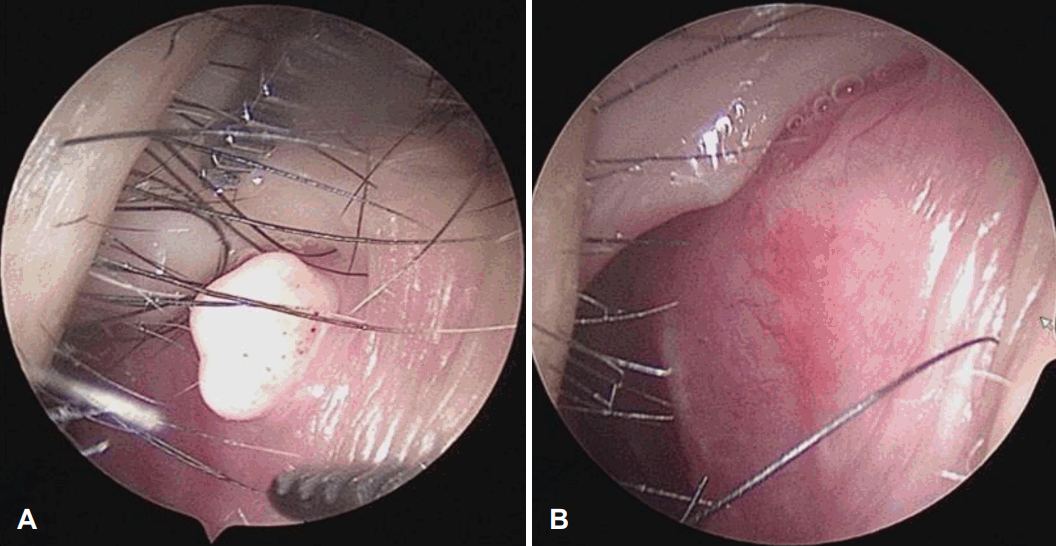
Preoperative and postoperative endoscopic images. A: Non-pedunculated whitish hard mass was firmly attached to the superior portion of the right nasal septal mucosa in the vicinity of the muco-epidermal junction. B: The surgical wound was well healed with no evidence of recurrence at postoperative 3 months.
병리조직검사 결과 조직의 내부는 섬유조직과 혈관 등의 중배엽 기원 조직들이 잘 발달된 소견을 보이고, 외부는 편평상피로 둘러싸여 있는 양상을 보여 전형적인 섬유상피용종으로 진단되었다(Fig. 2). 환자는 수술 후 3개월까지 외래 추적 관찰을 시행한 결과 재발 소견은 보이지 않았다(Fig. 1B).
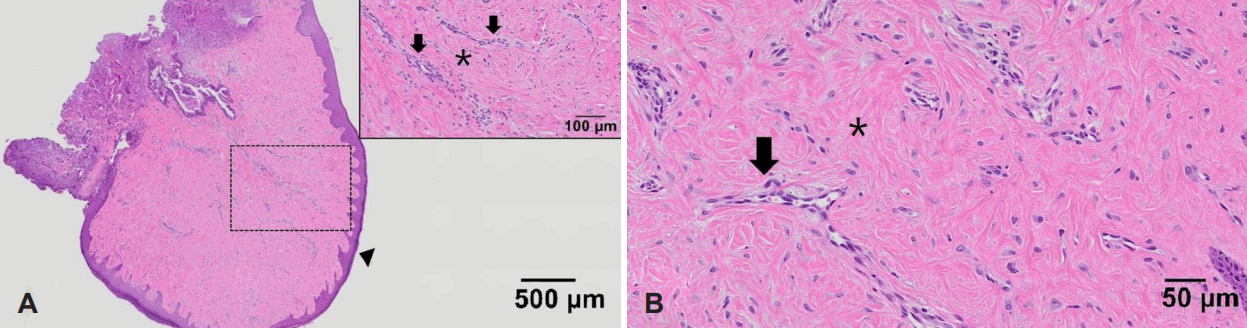
The microphotographs of the excised mass (hematoxylin and eosin staining). A: The stroma of the tumor is covered with squamous epithelium (arrowhead; ×40). The dotted rectangle from the mass is magnified at top-right corner of (×200). B: The stroma contains abundant fine collagen fibers (asterisk) and blood vessels (arrows), which are more evident in the further magnified microphotograph (×400).
고 찰
섬유상피용종은 피부의 중배엽 기원의 양성 결체 조직 종양으로 피부 쥐젖, 유경 연성섬유종으로 불린다. 피부 이외의 발생 부위로 위장관계, 요로계, 호흡기계에서 관찰되기도 한다[6]. 섬유상피용종이 상기도에서 발생하는 경우는 흔하지 않으나 인후두 부위에서 발생한 증례들이 보고된 바가 있다[7]. 육안적으로 섬유상피용종은 크기는 다양하나, 표면은 매끄럽고, 경계가 명확하다. 양성종양으로 대부분 특별한 증상을 유발하지는 않지만 출혈, 폐색, 통증 등을 유발할 수 있으며 주로 미용적 목적으로 치료가 요구된다.
섬유상피용종의 발생원인에 대해 여러 의견들이 있지만 아직 명확한 발병 기전이 밝혀지지는 않았다. 선천성 요인도 있다고 하나, 주로 후천적으로 만성적인 자극, 외상, 또는 알러지 반응에 의한다고 알려져 있다[4,7]. 비강 외의 위치에서 발생하는 섬유상피용종의 경우, 연기 흡인, 에스트로겐 호르몬에 의해 유발된다는 보고들도 있다[8,9]. 섬유상피용종은 비강 내에서는 매우 드물게 발생하며 원인과 발병 기전에 대해 알려진 것이 없다. 본 증례의 경우에도 병력에서 뚜렷한 발병 요인을 찾을 수는 없었으나, 계절성 비염 증상으로 반복적으로 코를 푸는 행위가 비중격에 만성적인 자극으로 작용했을 가능성이 있다.
섬유상피용종을 진단하기 위해선 먼저 비강 내에서 발생 가능한 다른 병변들과의 감별이 필요하며 병리조직학적 진단이 필수적이다. 섬유상피용종의 경우 병리조직학적 소견상 표면은 편평상피로 구성되어 있고, 내부는 주로 지방조직과 혈관조직 등의 중배엽 기원의 조직들로 구성되며 여러 기질세포들도 발견될 수 있다. 기질세포는 단핵성 및 다핵성 세포, 비전형적인 세포 등으로 구성되며 국소침습, 유사 분열성 등 악성 소견은 관찰되지 않는다[10]. 감별해야 될 비강 내 종물로 외성장유두종(exophytic papilloma), 염증성비용종, 호흡상피성 선종모양 과오종(respiratory epithelial adenomatoid hamartoma) 등이 있으며 병리조직학적 검사가 진단에 필수적이다[2].
기존 문헌에 따르면 비강 내 섬유상피용종의 경우 환자들은 코막힘, 간헐적 코피, 콧물 등의 증상을 동반할 수 있으며 절제술 시행 후 증상들은 모두 호전되었다. 또한, 수술적 절제 이후 재발된 사례는 없었다[1]. 비강 내에서 발생한 섬유상피용종의 경우 악성화가 발생한 증례가 보고된 바는 없었지만 피부에서 기원한 섬유상피용종의 경우 악성화된 증례가 보고된 바가 있다[11]. 따라서, 비강 내에서 발생한 섬유상피용종의 경우에도 완전한 수술적 절제를 시행하고 병리조직학적 검사를 시행하여 악성 여부를 확인하는 것이 필요할 것으로 생각된다.
Acknowledgements
None
Notes
Author Contribution
Conceptualization: Sang-Wook Kim. Data curation: Tae-Hun Lee. Formal analysis: Woohyen Jin, Sang-Wook Kim. Investigation: Woohyen Jin, Chang Hyeon Lee. Methodology: Tae-Hun Lee. Project administration: Sang-Wook Kim. Resources: Woohyen Jin, Tae-Hun Lee, Chang Hyeon Lee. Software: Chang Hyeon Lee. Supervision: Sang-Wook Kim. Validation: Woohyen Jin, Tae-Hun Lee. Visualization: Woohyen Jin, Chang Hyeon Lee. Writing—original draft: Woohyen Jin. Writing—review & editing: Woohyen Jin, Tae-Hun Lee, Sang-Wook Kim.