편측절골술을 통한 심한 외상성 사비의 교정 2예
Two Cases of Unilateral Osteotomies for Traumatic Severely Deviated Noses
Article information

Trans Abstract
A severely deviated nose accompanied with trauma is difficult to correct, but osteotomy, a common surgical technique, can correct such deviation. This study aims to evaluate the usefulness of unilateral osteotomy, a technique for correcting a severely deviated nose driven by trauma. Two patients, both with deviated nose caused by trauma but one with a C-shaped nose and another with a reverse C-shaped nose, underwent surgery. Cartilage and bone humps were removed by a knife and a rubin osteotome, respectively. Unilateral osteotomy was performed on the convex area to correct the crooked nose and camouflage dorsal graft was used to treat the lowered dorsum. No postoperative complications were reported during the follow-up periods. Both patients were satisfied with the outcomes. This paper presents successful cases of unilateral osteotomy and suggests this treatment as an effective way of correcting deviated nose caused by trauma.
Introduction
The crooked nose, defined as the deviation of the nasal pyramid from the midsagittal plane, is a major clinical condition requiring rhinoplasty [1]. Despite the fact that various methods for correction have introduced, recurrence rates are still high [2]. Osteotomy is a common treatment; nevertheless, it may cause soft tissue trauma. Major sequelae, accompanied by osteotomy, are edema and ecchymosis. Procedures of osteotomy should be precise and rigorous to reduce the possibility of morphological and functional complications [3]. Constructing subperiosteal tunnels prior to osteotomy was suggested to mitigate sequelae; however, the effect has yet been controversial [4]. We hypothesize that correcting traumatic deviated nose using unilateral osteotomy would be effective for rapid recovery and alleviation of morbidity. In spite of many studies on unilateral osteotomy for a severely deviated nose, this paper fills out the gap of repositioning a crooked nose induced by trauma. Therefore, we present two successful cases on hump resection and camouflage graft to emphasize the effectiveness of unilateral osteotomy. This study, covering a patient case report, was exempted from review by the Institutional Review Board of Kangwon National University Hospital (2022-08-006).
Case
Case 1
A 51-year-old male, suffering from nasal obstruction and external deviation, visited our clinic. His occupation was a police officer and was injured a decade ago while arresting a person who committed a crime. In the frontal view, the patient had a C-shape dorsal deformity, and the nasal septum was severely deviated to the right. Nasal hump was observed in bony-cartilage junction of dorsum from the lateral view and bulbous nasal tip also found in the basal view. To treat the patient’s condition, we planned to perform septorhinoplasty under general anesthesia using an open-approach technique. An inverted-V columellar and marginal incision were conducted. From the incision, the flap and the roof of cartilage were raised and dorsal skeleton was fully exposed. Since the anterior septal angle was exposed, we harvested septal cartilage and remained L-strut by performing septoplasty. Cartilage and bone humps were removed using a knife and rubin osteotome, respectively. Remaining rough surface of nasal dorsum was trimmed by rasp. In the process of eliminating hump, the concave side of the hump was more excised than convex side to sustain the symmetry after the midline shift. Since an open roof, created by humpectomy, helped meeting the lateral osteotomy, medial osteotomy was less required. Unilateral osteotomy was applied to the convex side of bone, and then movable convex side of pyramidal bone and septum were pushed toward the midline. After the procedures, camouflage dorsal graft Tutoplast® (Tutogen Medical GmbH, Neunkirchen am Brand, Germany) was implemented to restore the lowered dorsal height resulted from the hump removal (Fig. 1). To straighten, a spreader graft was placed on the concave side of dorsal cartilage using harvested cartilage. Septal extension and cap grafts were also performed with harvested cartilage to correct the bulbous nose. The improvements of the curvature of the nasal dorsum and overall contour are observed from the facial photograph of the patient taken a month after the operation, compared to the presurgery photograph (Fig. 2).
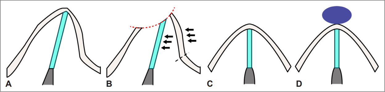
Schematic view of unilateral osteotomy. A: Preoperative view, one side of the nasal pyramid is concave, and the other is convex. B: Concave side of the hump was more excised than convex side (red dot line). Unilateral osteotomy was applied to the convex side of bone (black dot line), then was pushed toward midline (black arrow). C: Lowered dorsum height after procedure. D: Camouflage dorsal graft Tutoplast® (Tutogen Medical GmbH) was implemented.
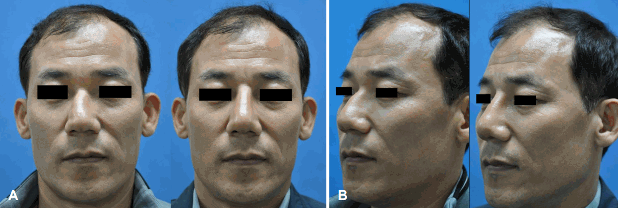
Perioperative facial photos. A: In preoperative image, there are C-shape deviation and dorsal deformity. B: Postoperative 4 weeks’ photograph shows correction of deviated nose with unilateral osteotomy.
Case 2
A 62-year-old male visited our clinic with symptoms of nasal obstruction and external nose deformities. The symptoms appeared after the bicycle accident that he had undergone three years ago. The patient’s nasal dorsum was deviated in “reverse C” shape, and saddle deformity was noted at keystone area (Fig. 3). A nasal cavity exam suggested that deviation of septum to right side would cause symptom of nasal obstruction. In order to correct the patient’s problem, we decided to perform septorhinoplasty. The operation was performed in a similar way to case 1. After conducting external approach with an inverted-V columellar and marginal incisions, a flap was lifted along the superficial muscular aponeurotic system layer. Septoplasty was performed to set deviated nasal septum, and cartilage was harvested afterwards. Then, dorsal hump was resected asymmetrically and medial osteotomy was performed for severely deviated nasal dorsum, in line with case 1. Unilateral osteotomy was applied along the nasofacial groove of the convex side. A Tutoplast® was inserted to the nasal dorsum as a camouflage graft to recover for lowered nasal dorsum. Additional spreader and septal extension grafts were used to straighten deviated dorsal cartilage portion and to increase tip projection, respectively. Preoperative and postoperative photographs show aesthetic changes in the patient’s nose (Fig. 4).
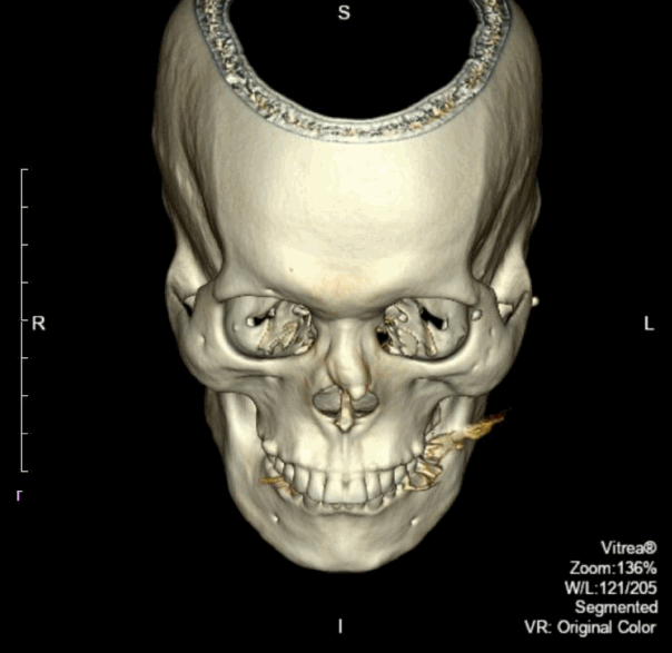
A representative 3-dimensional facial CT image (case 2). The nasal dorsum was deviated in reverse C-shape and a saddle deformity was placed at the keystone area.
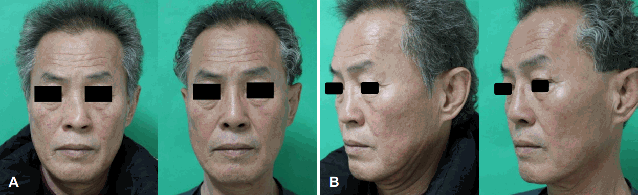
Perioperative facial photos. A: Reverse C-shape deviation and saddle deformity are shown on the preoperative image. B: A deviated nose corrected with unilateral osteotomy in postoperative 4 months’ photo.
Discussion
The deviated nose is defined as clinical conditions involving deviation of the nasal pyramid from the facial midsagittal plane [1]. This crooked nose is associated with bony or septal deformity, or both [5]. A deviated nose may appear congenitally or it may be acquired secondary like trauma. To recover these deviated nose, accurate diagnosis and abundant anatomic knowledge are required.
Osteotomy is an essential technique to correct a deviated nose in rhinoplasty. The success of rhinoplasty is closely related to making the nasal bony frame and soft tissue into the desired shape. Therefore, osteotomy is absolutely indispensable achieving aesthetic and functional outcomes, while satisfying these requirements simultaneously [6,7]. This surgical technique corrects open deformity after hump resection and narrows nasal pyramid, resected asymmetric region and made scoliotic portion to straight [8].
There are several methods for lateral osteotomy, which are primarily used, but no technique has adopted that minimizes side effects with preserving the effectiveness. Lateral osteotomy is dangerous surgical technique that has some side effects like tissue edema, ecchymosis, open roof deformity and airway stenosis [9,10]. Repetitive osteotomies may reduce postoperative stability and raise higher risk of adverse effects, especially on Asian noses. Asians have a low and flat bridge with a thick and small bony structure [11]. Therefore, we suggest unilateral osteotomy as a way of alleviating these side effects.
Sohn, et al. [12] reported 9 cases of correcting the deviated nose using unilateral osteotomy [12]. 5 cases were about C- or reverse C-shape, similar to our cases. Unilateral osteotomy was performed on a convex site, and a spreader graft and extension graft were used for nasal tip and nasal septum reconstruction. All of the surgical results were satisfactory, and none of the patients’ conditions has been worsened after the operation.
Unlike previous studies, unilateral osteotomy was performed focusing on a severely crooked nose that was accompanied by functional problems due to trauma. we focused on minimizing the invasiveness as a side effect while correcting a deviated nose with unilateral osteotomy, even in the patients had a severe deviated nose (C-shape, reverse C-shape) due to trauma. In the process of humpectomy, we removed hump asymmetrically to sustain the symmetry after the midline shift. After dorsal hump resection, we corrected the crooked nose by unilateral osteotomy and narrowing at convex portion, and augmented the nose by applying a camouflage graft.
In the case 1, C shape deviation recovered in straight shape in frontal view and in the oblique photograph, shown from the comparison of photographs. It confirmed that the rhinion area straightened due to hump resection and augmentation on the nasal dorsum side. The nasal tip was sharpened by camouflage graft, and the overall shape of the nose has become smooth (Fig. 2).
In case 2, the correction of reverse-C shape deviation in the frontal view is observed by the contrast of pre-operate and post-operate photos. In the oblique view, the contour of nose is straightened by nasal dorsum augmentation. The overall nasal height was elevated and nasolabial angle as well as nasofacial angle was increased (Fig. 4).
After the operation, the patients were satisfied with results, showing no complaints. The side effects, such as nasal obstruction, ecchymosis or saddle nose, and narrowing nasal valve, are not detected from the follow-up.
These two cases are too specific to represent unilateral osteotomy as the general or the only way of correcting traumatic deviated nose. Therefore, this treatment requires the further research to set the generally accepted standard on suitability for patients in multiple criteria. Despite of extensive literatures about unilateral osteotomy, further studies are required for the correction of a severely deviated nose generated by trauma. Our two cases would contribute by filling this gap.
Acknowledgements
None
Notes
Author contributions
Conceptualization: Woo Hyun Lee. Formal analysis: Woo Hyun Lee. Supervision: Woo Hyun Lee. Visualization: Shin-woo Kim, Yoon Heo. Writing—original draft: Shin-woo Kim. Writing—review & editing: Shin-woo Kim, Yoon Heo, Ye-Sol Jung.