선천성 유양동 진주종을 포함한 세 가지 외배엽 기원의 선천성 기형 1예
A Case of Three Ectodermal Origin Congenital Anomalies Including Congenital Mastoid Cholesteatoma
Article information
Trans Abstract
There are several congenital anomalies of the mastoid and external ear. The external ear and mastoid form during the developmental process through the differentiation of ectoderm and mesoderm, and anomalies can arise at various stages. While most congenital anomalies can be easily diagnosed through physical examination, some cases, such as congenital mastoid cholesteatoma, may delay diagnoses. We present a successfully treated case that involved the removal of three simultaneous congenital anomalies of ectodermal origin, which included preauricular fistula, congenital mastoid cholesteatoma, and postauricular epidermal cyst, all found on one ear in a single patient.
서 론
귀와 유양동에서는 발생 과정에서 다양한 선천 기형이 생길 수 있다. 특히 외이와 중이는 발생학적으로 서로 관련이 있는 경우가 많아 외이의 기형이 있는 경우 외이도나 중이의 동반 기형이 나타나기도 한다[1]. 중이에서는 대표적으로 외배엽 기원의 선천성 진주종, 중배엽 기원의 선천성 이소골기형이 발생할 수 있으며, 외이에서는 소이증, 이수 기형, 전이개 누공 같은 이개기형과 외이도 협착, 외이도 누공과 같은 외이도 기형과 같은 외배엽 기원의 기형이 발생할 수 있다. 귀에 나타나는 기형들은 대부분 단일 기형으로 나타나나 복합 기형이 존재하는 경우도 있다. 최근 저자들은 외이와 유양동에서 전이개누공, 선천성 유양동 진주종, 선천성 표피낭종이 함께 발견된 증례를 경험하였으며, 수술적 치료를 시행하여 문헌고찰과 함께 보고하는 바이다.
증 례
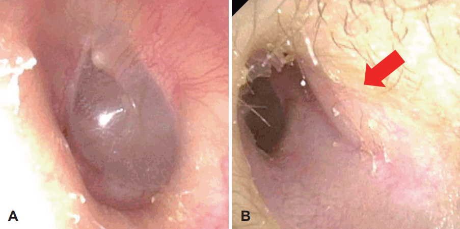
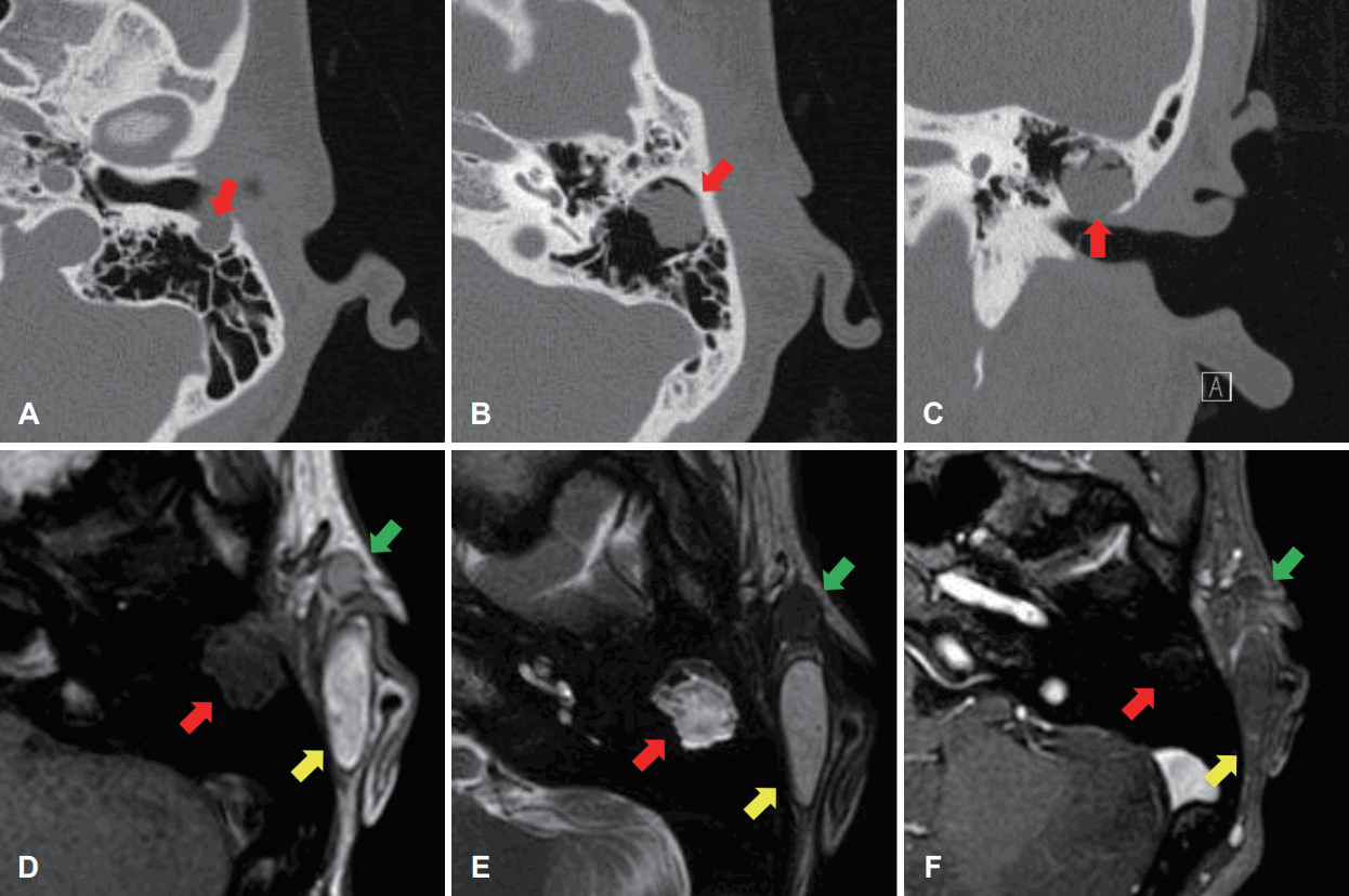
17세 여자 환자가 좌측 전이개 누공의 분비물을 주소로 수술적 치료를 권유받아 내원하였다. 가와사키 병을 앓았던 것 외에 특이 과거력은 없었으며, 귀와 관련된 수술력은 없었다. 신체진찰상 좌측 전이개 누공과 함께 후이개 상방의 2×2 cm 크기의 종물이 관찰되었다. 양측 고막은 정상이었다. 전이개 누공과 이개 후상방 종물의 평가를 위해 측두골 컴퓨터단층촬영을 시행하였다. 좌측의 전이개 누공, 후이개 표피낭종과 함께 유양동 내 진주종으로 의심되는 병변이 확인되었고, 외이도 후벽의 골파괴 소견이 함께 관찰되었다. 재시행한 귀 내시경 검사상 외이도 후벽의 일부에서 골결손부를 확인할 수 있었다(Fig. 1). 함께 시행한 측두골 자기공명영상 검사에서 유양동 종물은 T1에서 저신호강도, T2에서 고신호강도를 보여 선청성 진주종을 의심할 수 있었다(Fig. 2) [2]. 청력검사 및 고실 임피던스 검사에서 특이소견은 발견되지 않았으며, 이명, 난청, 이루, 어지럼증 등 동반되는 증상은 없었다. 영상 검사에서 유양동 진주종, 전이개 누공 및 후이개 표피낭종, 세 병변 간의 연결은 없었다.

Endoscopic images of left external auditory canal. A: Tympanic membrane. B: The posterior wall of the external auditory canal is bulging (red arrow), and bony destruction is observed.

Radiologic images of three congenital anomalies of ear. A: Bony destruction of posterior wall of the external auditory canal is identified on the axial view of the CT (red arrow). B: Isolated mastoid cholesteatoma (red arrow). C: Bony destruction of the external auditory canal wall is observed (red arrow). D: Low signal isolated mastoid cholesteatoma (red arrow), a low signal preauricular cyst (green arrow), and a high signal post-auricular epidermal cyst (yellow arrow) were identified on the T1 axial view. E: High signal isolated mastoid cholesteatoma (red arrow), low signal preauricular cyst (green arrow), and low signal post-auricular epidermal cyst (yellow arrow) was noted on the T2 axial view. F: All three lesions (epidermal cyst in yellow arrow, mastoid cholesteatoma in red arrow, preauricular cyst in green yellow) were characterized by low signal intensity on the gadolinium-enhanced view.
세 가지 선천성 종물에 대하여 수술적 절제를 계획하였다. 전이개 누공은 메틸렌 블루를 이용하여 전이개강 내를 표지하고 타원 모양의 절개를 통해 이륜 연골까지 이어지는 피하낭을 완전히 절제하였다. 이어 후이개구를 따라 절개선을 넣고, 피하에 있던 경계가 명확한 후이개 표피낭종을 제거하였다. 마지막으로 후이개구 절개선 부위를 따라 피판을 거상한 뒤, T자 형으로 골막을 절개하여 유양동을 노출하였다. 유양동 표면의 골파괴소견은 없었다. 절단형 버어를 이용하여 유양동 삭개술을 시행하였으며, 개방형 유양동 진주종을 확인할 수 있었다. 진주종으로 인한 외이도 후벽의 골 파괴 부위를 확인하였고, 유양동 진주종을 완전히 제거한 뒤 진주종 부착부 및 골파괴 부위를 다이아몬드 버어로 정리하였다. 외이도 후벽은 연골편과 측두근 근막을 이용하여 재건하였다(Fig. 3). 수술 소견상 각 병변의 경계는 명확하고, 연결성은 찾을 수 없었다. 세 부위의 병리 결과는 각각 전이개 누공, 표피낭종, 진주종으로 보고되었다(Fig. 4). 술후 3일에 이개 부종, 외이도 부종 등 연골막염 의심 소견이 있어 외이도 패킹을 시행하였으며, 항생제 사용 후 증상 호전되어 술후 11일에 합병증 없이 퇴원하였다. 술후 1개월 시행한 신체검사상 수술부위와 고실 모두 이상소견 없이 관찰되었다. 보강한 외이도 벽도 정상적으로 확인되었다. 재발없이 추적관찰 중으로, 향후 유양동 진주종 재발 여부를 확인하기 위해 측두골 컴퓨터단층촬영을 계획 중이다.

Intra-operative images. A: Preauricular cyst. B: Post-auricular epidermal cyst. C: Isolated mastoid cholesteatoma (green arrow) and bony destruction of the external auditory canal wall (yellow arrow). D: After reconstruction of external auditory canal posterior wall using conchal cartilage and temporalis muscle fascia (yellow arrow).
고 찰
귀는 발생 과정 중 여러 단계에서 선천성 기형이 발생할 수 있다. 불충분한 발생 및 융합, 과증식, 불충분한 증식 등이 원인으로 생각되며, 유전적인 요인이 보고되기도 한다[3]. 외이는 태생 4주 경 제1새궁과 제2새궁의 간엽 조직이 응축되고 태생 6주 경 발생한 6개의 이개융기들이 융합하고 분리되면서 발생 20주 경 성인 이개 모양을 형성한다. 제1새궁과 제2 새궁은 이개, 중이, 내이, 안면신경, 하악골, 상악골, 설골 등의 구조물로 발생한다[4]. 외이도는 태생 4주경 제1새열이 넓어지고 외배엽이 증식하면서 관의 형태를 띄며 발생하고, 태생 8주경 외배엽 세포들이 본격적으로 증식하기 시작하여 외이도 전을 형성한다. 이개 발생 과정 중 제1새궁과 제2새궁의 표피 이동(epithelial migration)에 이상이 있는 경우 전이개 누공과 선천성 진주종이 동시에 발생할 수 있다[5].
일반적인 전이개 누공 환자에서는 영상검사를 시행하지 않으나, 본 증례에서는 후이개 종물과의 연결성 평가를 위해 영상검사를 시행하였다. 영상검사에서 동측의 선천성 유양동 진주종, 전이개 누공과 연결이 없는 후이개 표피 낭종이 확인되었고, 진주종으로 인한 외이도의 결손이 보였다. 환자는 특이 가족력은 없었으며, 다른 기형이나 장애는 없었다. 수술 소견상 세 병변은 서로 연결되지 않은 형태였으며, 성공적으로 제거되었다. 전이개 누공은 태생 6주의 제1새궁, 제2새궁에서 발생한 원시이개 중 6번 이개융기의 불완전한 병합에 의해서 발생하는 것으로 알려져 있다[6]. 대개 전이개 누공의 작은 구멍은 이륜의 전단부에 위치하고 이륜의 후상부나 이주, 이수에서도 발견되며, 낭종은 다양한 방향으로 트랙을 형성한다. 상염색체 우성 유전을 보이는 Branchio-oto-renal 증후군은 전이개 누공, 경부 누공과 같은 새열 기형과 함께 외이, 중이, 내이의 구조적인 결함과 감각신경성, 전도성, 혼합성 난청이나 신장 이상이 동반된다[7]. 전이개 누공과 선천성 중이 진주종이 동시에 발생한 증례들이 보고되었으나, 본 증례와 같이 전이개 누공과 함께 선천성 유양동 진주종, 표피낭종이 보고된 바는 없었다[5,8]. 전이개 누공 증례에서 비특이적인 위치에 누공이 있거나, 다른 기형이 의심되는 경우, 병변의 범위의 평가가 필요한 경우는 영상검사가 필요할 수 있다.
선천성 진주종은 측두골의 여러 부위에서 발생할 수 있지만, 유양동에 국한된 선천성 진주종은 매우 드물게 보고된다[9]. 선천성 진주종은 표피낭종의 하나로 외배엽 기원의 선천성 질환이다. 발생 기전은 표피양 형성설(epidermoid formation) [10], 상피 이동설(epithelial migration theory) [11], 고막 내함 및 유착설(repeated retraction and adhesion) [12], 상피화 생설(squamous metaplasia) [13] 등의 다양한 가설로 설명한다.
유양동에 국한된 선천성 진주종은 1) 이전 귀 수술력이 없고 이루가 없으며 정상 고막을 지니며 2) 영상학적으로 중이나 유돌동구, 상고실의 침범이 확인되지 않고, 수술 중 해당 부위의 진주종 소견이 없는 경우 진단할 수 있다[14]. 일반적인 선천성 중이 진주종은 평균 6.5세에 발견되며, 삼출성 중이염 진단시 우연히 발견되는 경우가 많다. 반면 선천성 유양동 진주종은 평균 47세에 발견되며, 주로 측두부에 국한된 통증과 부종을 호소하며, 경부 통증, 청력 저하, 급성 유양돌기염 등으로 내원한다[9]. 선천성 유양동 진주종은 유양동 피질골의 파괴로 인한 감염이 될 수 있기 때문에 귀 주위 통증이 있는 경우는 적극적인 영상 검사를 통해 조기 진단하여 심각한 합병증을 예방해야 한다[14].
본 증례는 우연히 발견된 선천성 유양동 진주종으로 환자가 호소하는 특별한 증상은 없었다. 수술 소견상 개방형 선천성 진주종으로 확인되었다. 개방형 선천성 진주종은 폐쇄형 진주종에 비하여 병의 진행이나 증상이 심해 골 파괴, 이소골 미란, 미로 누공, 안면신경관 미란 등이 흔히 나타나며, 수술 후 잔존 진주종의 빈도가 높다[15]. 본 증례에서 함기화가 잘된 유양동에 넓게 퍼져 있는 진주종을 성공적으로 제거하였으며, 잔존 진주종을 방지하기 위해 부착 부 및 골 파괴가 있었던 유양동 부위를 다이아몬드 버어로 절삭하였다.
본 증례를 통해 임상에서 흔히 볼 수 있는 전이개 누공 환자에서 면밀한 신체진찰의 중요성과 동반 기형이 있을 때 중이와 외이, 경부에 대한 영상검사가 필요할 수 있음을 알 수 있었다. 저자들은 전이개 누공과 동반된 선천성 유양동 진주종의 발생학적 의의를 문헌 고찰과 함께 보고하는 바이다.
Acknowledgements
This research was supported by a grant from the Korea Health Technology R&D Project through the Korea Health Industry Development Institute (KHIDI), funded by the Ministry of Health & Welfare, Republic of Korea (grant number: HI21C1574).
Notes
Author contributions
Conceptualization: Jae Ho Chung. Data curation: Woogeun Seo, Junsung Bahn. Investigation: Minju Baek. Project administration: Jae Ho Chung. Resources: Jae Ho Chung. Supervision: Jae Ho Chung. Visualization: Minju Baek, Junsung Bahn. Writing—original draft: Woogeun Seo. Writing—review & editing: Jae Ho Chung.

