인두피부누공에서 진공을 이용한 관내 음압 드레싱의 효과
Usefulness of Endoluminal Vacuum-Assisted Closure Therapy for Pharyngocutaneous Fistula
Article information
Trans Abstract
Pharyngocutaneous fistula (PCF) is a communication created between the pharynx and skin of the neck associated with flap reconstruction or primary closure of a pharyngeal defect. In patients who underwent laryngectomy and pharyngectomy, salivary accumulation in the deep neck space through the pharyngeal dehiscence can cause life-threatening complications, among which is carotid artery blow-out. Broad-spectrum antibiotics, compressive dressing, and artificial nutrition are usually applied first. Many closing methods have been studied, both surgically (direct repair or using flaps) and non-surgically (negative pressure therapy, hyperbaric oxygen therapy, or botulinum toxin injection). The use of a vacuum-assisted closure system placed endoluminally or externally has been recently reported to treat any leaks in the upper gastrointestinal tract. Here, we report a successfully-treated PCF using the endoluminal vacuum-assisted closure system.
서 론
인두피부누공은 인두와 경부 피부와의 통로가 생긴 것을 의미한다. 후두 절제술이나 인두 절제술 후 봉합된 인두에 틈이 생기면 침 등이 경부로 흘러나올 수 있는데, 경동맥 파열처럼 심각한 결과를 초래할 수 있는 잠재적으로 위험한 합병증이다[1,2].
인두피부누공의 치료는 보존적인 요법을 시행하면서 해결이 될 때까지 기다리거나 조기에 적극적 치료를 시행할 수도 있다. 적극적인 치료로는 유경 혹은 유리 피판을 이용하거나 내시경적으로 수복하는 관혈적 수술적 방법과 고압산소요법, 보톡스 주입법, 그리고 진공을 이용한 음압치료와 같은 비수술적인 치료가 있다[3-5].
최근 직장 절제술 후 열개와 식도 천공과 같은 위장관의 누출 때 진공을 이용한 치료가 보고되고 있다[6-9]. 인두피부누공 때에도 진공을 이용한 음압 드레싱을 적용하여 치료한 보고가 있다[10].
이에 본 연구자는 재건된 인두의 열개로 인한 인두피부누공 환자를 진공을 이용한 음압 드레싱을 인두 관내에 적용(endoluminal vacuum-assisted closure, EndoVAC)하여 치료하였기에 보고하고자 한다.
증 례
본 연구는 화순전남대학교병원 연구윤리심의위원회(Institutional Review Board)의 승인을 받았다(CNUHH-2023-159).
60세의 남자가 여러 차례 두경부에 편평세포암이 재발하여 수술을 받았다. 우측 조기 성문암으로 방사선 치료 후 3년 후에 같은 곳에 재발하여 상윤상 후두 부분 절제술 및 윤상설골후두개 고정술 시행받았고, 그 후 1년 5개월 만에 재발하여 후두 전적출술을 받았고, 그 후 1년 7개월 만에 경부식도 및 피부에 재발하여 식도 부분절제술 및 삼각흉근 피판 재건술을 받았고, 그 후 10개월 만에 식도에 재발하여 하인두 및 식도 전절제술 및 위장 견인 재건술을 받았다. 재발의 중간중간에는 항암제 치료도 받았다. 그러나, 여러 차례의 수술과 항암제 치료에도 불구하고 최근 구인두 후벽 하방, 즉 견인 재건된 위장과의 경계 부위에 재발이 확인되어 인두 및 견인된 위장의 부분 절제술 및 유경 대흉근 피판 재건술을 받았다.
수술 2일 후에 혼탁한 배액관이 관찰되었다. 경부 절개선을 손으로 눌렀을 때 혼탁한 액체가 흘러나오는 것이 확인되었다. 전산화단층촬영상 재건된 인두의 열개를 통한 심경부의 액체 저류가 확인되어 응급수술을 시행하였다. 개구기를 장착하고 인두벽의 열개를 확인하려 하였으나 노출이 잘 되지 않았다. 양측 경부를 절개했을 때 농과 함께 침이 확인되었으며, 배농술과 함께 배액관을 삽입한 뒤, 영양공급을 위한 공장루를 만들고 수술을 마쳤다.
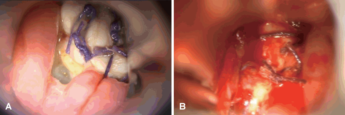
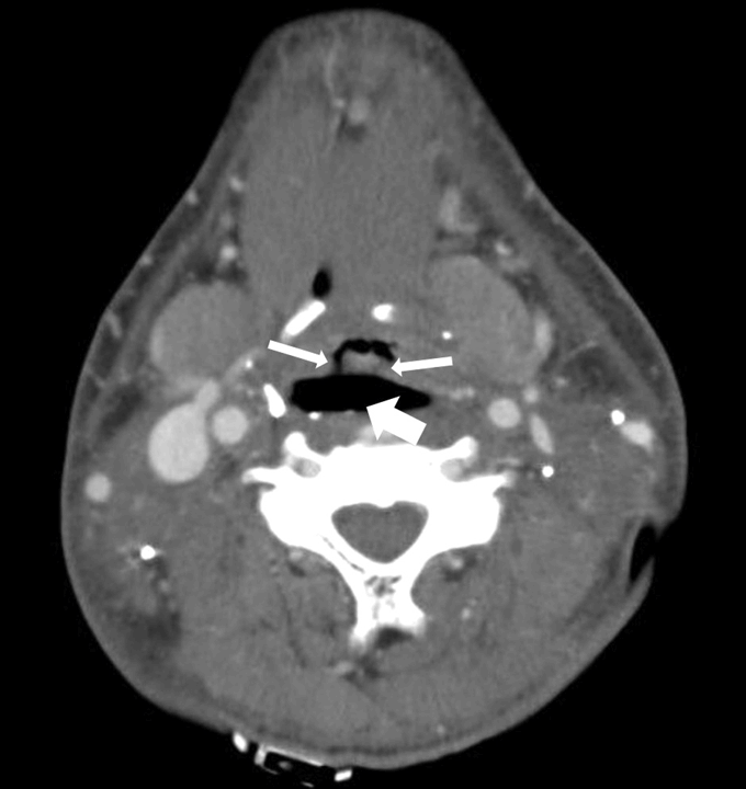
2일 후에 다시 전신마취를 시행하였다. 경구강을 통해 구인두 후벽과 재건된 대흉근 피판에 열개가 확인되었다(Fig. 1A). 피판의 혈류 상태는 양호하였으며, 열개는 잔존암 혹은 재발에 의한 것이 아님을 확인하였다. 인두경을 장착한 상태로 경구강을 통하여 내시경적 봉합을 시행하였다(Fig. 1B). 후인두의 사강(dead space)에 국소지혈제를 뿌려 사강을 줄이고 봉합을 안정화하려 하였다. 그러나 다음날 시행된 전산화단층촬영상 열개가 더 심화된 것이 확인되었다(Fig. 2). EndoVAC으로 열개를 해결하고 결국 인두봉합누공을 치료하고자 하였다.

The dehiscence was confirmed on the posterior oropharyngeal wall under suspension pharyngoscopy (A) and closed endoscopically intraorally (B).

The neck CT taken one day after the endoscopic repair for the anastomotic dehiscence demonstrates more aggravation of the dehiscence (thin arrows) and a large amount of air pocket in the retropharyngeal space (thick arrow) even though the effort of the repair.
식도 천공에 대한 EndoVAC 삽입은 문헌보고에 따르면 전신마취하에서 시행되었으나[9], 본 증례는 열개 부위가 인두에 국한되어 있어 국소마취로 수행이 가능할 것 같아 시도해 보았다. 먼저 리도카인액으로 구강, 구인두 및 비강을 국소 마취하였다. 14 Fr. 크기의 Levin 튜브를 한쪽 코로 통과시켜 입으로 빼낸 다음 구멍이 뚤린 부위까지 잘라내었다(Fig. 3A). 2차 치유를 목적으로 삼출액이 많은 창상의 보호를 위해서 사용하는 친수성 폼제 피복재인 스펀지(V.A.C. Granufoam Dressing; KCI Manufacturing, Athlone, Ireland)를 인두관 내를 충분히 채울 정도의 크기로 디자인하였다(Fig. 3B). 스펀지 한쪽 끝의 가운데 부분에 가위를 이용하여 Levin 튜브가 들어갈 정도로 구멍을 내고 겸자로 반대편에 완벽히 관통하지 않을 정도의 끝부분까지 구멍을 연장시켜 뚫었다(Fig. 3C). Levin 튜브와 스펀지가 고정되도록 두 군데를 실로 고정하였다(Fig. 3D). 굴곡형 후두내시경 가이드 하에 고정이 된 스펀지를 겸자로 잡아서 열개 부위의 인두에 위치되도록 구강을 통해 넣고(Fig. 4A) 제대로 삽입되었는지 확인하였다(Fig. 4B).

General outline of the endoluminal vacuum-assisted closure insertion. A: 14 Fr.-sized Levin tube is passed out to the mouth through the one nasal cavity and cut at the end of any hole. B: A hydrophilic sponge for secondary wound healing (V.A.C. Granufoam Dressing; KCI Manufacturing) is prepared and designed to be fitted into the intraluminal space of the pharynx. C: A hole is made into the middle of the one side of the sponge and advanced using clamps until the very end of the other side. D: The Levin tube and the sponge are anchored together with two stitches.

Endoscopy-guided endoluminal vacuum-assisted closure insertion. A: The sponge is grasped and inserted into the oropharynx at the point of dehiscence endoluminally under the guide of flexible laryngoscopy. B: The final position is confirmed by flexible laryngoscopy.
인두에 삽입된 스펀지를 진공 상태로 유지하기 위해 비강을 통해 나와 있는 Levin 튜브의 끝부분에 음압 상처 드레싱(KCI Medical Products LTD., Wimborne, Dorset, UK)을 적용하였다. 압력은 125 mm Hg로 유지하고 지속(continuous) 모드로 세팅하였으며, 환자가 이동을 할 때 쉽게 휴대할 수 있게 하였다. 간혹 환자가 입을 벌릴 때 순간적으로 양압 상태가 된 다음 음압으로 잘 환원되지 않으면 입원실 벽에 있는 흡인장치(wall suction)에 연결하여 음압으로 바로 환원될 수 있도록 하였으며, 만약 환자가 이동해야 하는 경우에는 입원실에 돌아왔을 때 바로 다시 연결하여 음압이 걸리도록 하였다.
이런 형태로 추후 6일 간격으로 스펀지를 2차례 교체하여 EndoVAC 치료를 지속하였다. 이 두 번의 교체는 환자의 편의와 인두 부위의 관찰, 그리고 스펀지 위치의 정확성을 기하기 위해 전신마취에서 시행하였다. 첫 번째 교체 후 스펀지가 삽입되었던 인두벽에는 열개가 사라지고 작은 크기의 다발성 건강한 육아조직이 형성되어 있는 것을 확인할 수 있었다(Fig. 5).

Small multiple healthy granulation tissue is formed on the nearly entire wall of the oropharynx with resolution of the dehiscence.
총 세 차례의 EndoVAC 치료 후에 전산화단층촬영을 통해 열개와 인두피부누공이 해결된 것을 확인하였다(Fig. 6). 스펀지를 구강을 통해 겸자로 제거하고 1주일이 지난 후 구강식이를 재개하면서 공장루는 제거하였고, 큰 문제없이 퇴원하였다.

CT taken during the third time of endoluminal vacuum-assisted closure therapy demonstrates the resolution of the dehiscence and retropharyngeal air pocket and remarkable maintenance of the luminal space of the oropharynx.
한 달이 지난 후 항암방사선 치료 동시요법을 받는 도중 인두협착이 발생하여 확장술을 시행하였고, 전과 같이 구강식이를 활발히 하고 있다.
고 찰
국내보고를 통한 인두피부누공의 원인으로는 수술 전에 받았던 방사선 치료[11,12], 병변이 광범위하여 후두 전적출술 때 인두재건이 필요했던 경우[11,13,14], 수술 전 낮은 알부민 수치[12,14], 수술 전 낮은 헤모글로빈 수치나 흡연의 과거력 혹은 기저질환의 동반[12] 등이 있다.
인두피부누공의 치료는 대략적으로 다음과 같다. 우선 누공이 확인되면 광범위 항생제 투여, 압박 드레싱, 그리고 인두를 우회하여 장이나 정맥을 통한 영양 공급을 시행한다[3]. 이렇게 보존적인 요법을 시행하면서 해결될 때까지 기다릴 수도 있고, 조기에 적극적 치료를 시행할 수도 있다. 적극적인 치료로는 유경 혹은 유리 피판을 이용하거나 내시경적으로 수복하는 관혈적 수술적 방법과 고압산소요법, 보톡스 주입법, 그리고 진공을 이용한 음압치료와 같은 비수술적인 치료가 있다[3-5].
폴리우레탄 스펀지를 통한 음압 폐쇄 요법은 표재성 상처를 치료하는 데 상당히 정형화 되어 있는 방법이다. 이것은 육아조직의 형성을 증대하고, 혈관성을 높이고 저류는 감소시킴으로써 상처 회복을 촉진시킨다. 두경부 영역에서는 여러 다양한 곳에서 높은 성공율을 보이며 적용되어 왔다.
관내(endoluminal space)에서의 적용은 주로 위장관, 즉 직장절제술 후 열개와 식도 천공과 같은 상황에서 이용되어 왔다[6-9]. 따라서 인두에서도 관내 적용이 충분히 가능하다고 예측할 수 있겠다. 인두봉합을 강화하는 과정에서 관내에 스펀지를 적용한 연구가 있었는데, Asher 등[15]은 12명 중 11명의 환자에서 성공적인 결과를 보고하였다. 그러나 이것은 인두봉합을 할 때 유경 혹은 유리피판을 같이 시행하였으며, 동시에 스펀지를 인두강 전체에 걸쳐 넣었던 경우였다.
인두피부누공 때에 진공을 이용한 음압 드레싱을 적용한 Steinbichler 등[10]의 연구는 경경부(transcervial), 즉 기관절개공 근처의 누공을 통해 스펀지를 넣어서 인두의 열개 근처까지 위치시키는 치료였다. 그들은 이것을 ‘outside-in technique’이라고 하였다. 이 방법만으로 6명의 환자 중 5명에서 성공적으로 누공을 해결하였으며 스펀지를 적용한 평균일은 18.5 (7-32)일이었고, 평균적으로 4 (1-9)번에 걸친 스펀지의 교체가 이루어졌다. 국내에서는 Park 등[6]이 갑상선 수술 도중에 생긴 식도 천공에서 진공을 이용한 음압 상처 드레싱을 적용하였다. 이들은 3-5일 간격으로 총 15번의 스펀지를 교체하였고 73일 만에 식도 천공이 해결되었다고 하였는데, 이것 역시 경경부, 즉 외부에서 스펀지를 적용하여 치료한 경우였다. 따라서 본 연구는 인두피부누공 때 추가적인 유경 혹은 유리피판의 수술 없이 관내로 스펀지를 적용하여 진공을 이용한 음압 치료만으로 어느 정도 성공적인 결과를 얻었기에 그 연구 가치가 있다고 하겠다.
본 증례와 같이 EndoVAC의 장점은 인두피부누공의 원인이 되는 인두의 열개 부위에 직접 적용을 할 수 있다는 것이다. 음압을 걸어 구강 내용물을 흡인하기 때문에 추가적인 타액 등의 구강 내용물이 심경부 공간에 저류되는 것을 방지할 수 있고, 위에서 역류된 내용물이 경부로 다시 들어가는 것도 차단할 수 있다. 전신마취를 할 수 없는 환자에서도 경우에 따라서는 가벼운 진정하에서 수행할 수 있으며, 피판 재건술 같은 크고 장기간의 시간이 필요한 환자에서 인두피부누공을 해결할 수 있는 대안이 될 수 있다. 술후 인두피부누공이 예상되는 환자에서 처음으로 인두를 재건할 때 EndoVAC을 예방 목적으로 삽입할 수도 있으며, 피판을 이용해서 인두를 재건하는 경우에도 피판 안에 EndoVAC을 미리 거치하여 예방할 수도 있다. 한쪽 비강으로는 EndoVAC을 넣고 다른 비강으로는 비위관을 넣어 이 곳을 통해 경장 영양공급을 동시에 시행할 수도 있다. 피판 재건술 같은 고도의 기술을 시행할 수 없는 의사들도 어느 정도 교육을 받으면 비교적 쉽게 수행할 수 있다.
다만 몇 가지 단점이 있을 수 있는데, 열개 부위가 큰 혈관에 가깝거나 혈관이 노출되어 있을 때는 그 사용이 추천되지 않는다. 또한 Levin 튜브가 코를 자극하여 통증을 유발하거나, 구인두에 오랫동안 거치된 스펀지가 인두 불편감을 초래할 수 있다. 제한된 공간에서 EndoVAC을 삽입하기 때문에 시야가 좁아서 열개 위치를 잘 파악한 후 넣어야 한다. 스펀지가 음압으로 인해 주변 조직과 유착되어 가벼운 견인으로는 쉽게 떨어지지 않거나 분리된 후 소량의 출혈이 생길 수도 있는데, 이 때는 식염수를 점적한 후 제거하면 된다. 스펀지를 장기간 거치할 수 없고, 삼출물이 시스템을 폐쇄할 수도 있기 때문에 문헌[10]에서는 2-4일 간격으로 스펀지를 교체할 것을 추천하고 있다. EndoVAC의 사용으로 그 발생율이 높지는 않으나 향후 내강이 협착될 수 있으며[10], 이 때는 확장술이나 스텐트 삽입술을 시행할 수 있다.
Acknowledgements
None
Notes
Author contributions
Conceptualization: Joon Kyoo Lee. Data curation: Hyeong Beom Lee, Juwon Hwang, Joon Kyoo Lee. Formal analysis: Joon Kyoo Lee. Investigation: Joon Kyoo Lee. Methodology: Joon Kyoo Lee. Project administration: Joon Kyoo Lee. Resources: all authors. Software: Hyeong Beom Lee. Supervision: Joon Kyoo Lee. Validation: Joon Kyoo Lee. Visualization: Gun Ju Kim. Writing—original draft: Hyeong Beom Lee, Joon Kyoo Lee. Writing—review & editing: Joon Kyoo Lee.
