 |
 |
AbstractNeuromyelitis optica (NMO) is an inflammatory demyelinating disease of the central nervous system, characterized by repeatedly invading the brain, optic nerve, or spinal cord. In 2004, when a disease-specific antibody (aquaporin-4-immunoglobulin G) was discovered in the serum of an optic neuromyelitis patient, it was thought to be a separated disease from multiple sclerosis. Since 2004, however, the concept of the disease has been expanded to include NMO spectrum disorder (NMOSD). We experienced a rare case of NMOSD in a patient who presented with left visual disturbance and underwent endoscopic sinus surgery because of acute fungal sinusitis. We report this case with a review of the literature.
IntroductionNeuromyelitis optica spectrum disorder (NMOSD) is a rare inflammatory disease against the aquaporin-4 (AQP4) water channel that most often affects the optic nerves and spinal cord [1]. Several attacks can cause severe neurological damage leading to optic neuritis and transverse myelitis [1]. Since the discovery of the AQP4 antibody (AQP4-Ab) biomarker, NMOSD has been regarded as a specific entity with pathological, radiological, and clinical features that are reliably different from those observed in multiple sclerosis (MS) [1]. New clinical criteria for NMOSD based on AQP4-Ab serostatus has improved diagnostic accuracy and reduced the delay in diagnosis [1].
Invasive fungal sinusitis (IFS), is a potentially fatal disease, often presenting as an acute or subacute, painful orbital apex syndrome, with blindness and ophthalmoplegia [2]. Common general symptoms of IFS include nasal obstruction, rhinorrhea, bleeding, fever and facial pain. Once in the orbit, ocular symptoms include decreased visual acuity, diplopia, swelling, ocular pain, limitation of ocular movement, trigeminal hypoesthesia or hyperalgesia, proptosis and chemosis [3]. Presenting signs and symptoms of IFS commonly overlap with other diseases such as infection, allergy, autoimmune and neoplastic disease. Differential diagnosis are essential for proper treatment.
CaseA 69-years-old male visited to the department of ophthalmology, university hospital with complaining left visual disturbance and orbital swelling. He had no symptom associated to nose such as nasal obstruction, rhinorrhea or anosmia. The family histories and past histories were negative for significant disorders. The patient checked fundus photography, visual field exam and oribit MRI in the out patient department of ophthalmology. Orbit MRI showed left ethmoid fungal sinusitis with optic neuritis, the patient was transferred to otorhinolaryngology department. Paranasal sinus (PNS) CT with thin section enhanced and laboratory examination were checked. PNS CT showed left ethmoid fungal sinusitis with mild enhancing left cavernous sinus. On sinus endoscopic examination, we could find mucoid rhinorrhea drained from left osteo-meatus unit site. We prepared for an emergency endoscopic sinus surgery (Fig. 1).
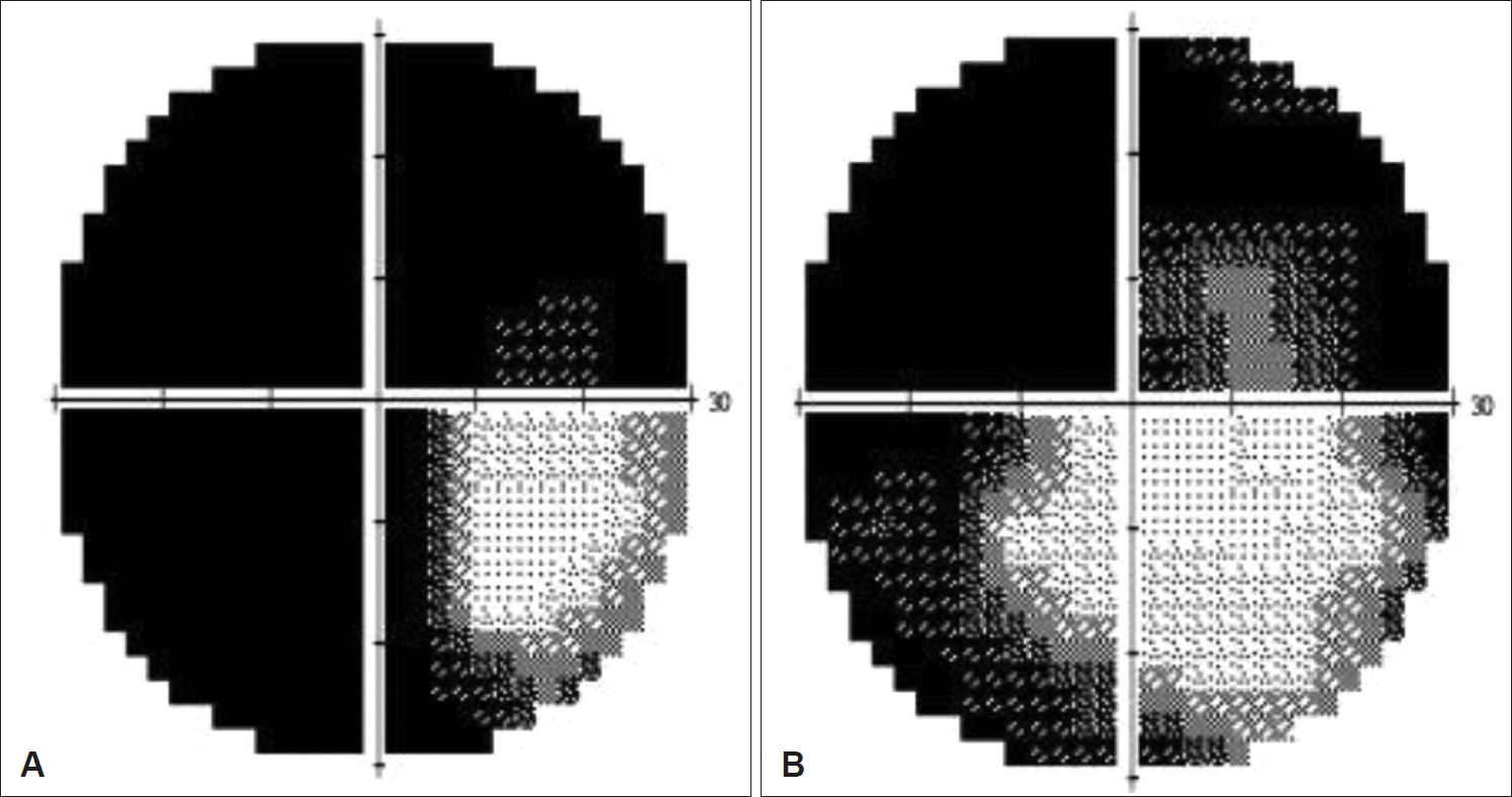
Both middle meatal antrostomy, left anterior, and posterior ethmoidectomy were performed. Fungal balls were identified in the left ethmoid sinus, followed by biopsy and culture. Severe mucosal swelling and mucoprulent rhinorrhea were observed by sinus endoscope in the operative findings, and the department of infectious medicine consultation was performed for using an antifungal agent for orbital complications. (1-3)-β-D-glucan test resulted positive. Aspergillus infestation was confirmed by biopsy, and voriconazole was used after transfer to the department of infectious medicine. The patient gradually recovered visual disturbance after surgery. In the visual field exam, a recovery was observed from 12% to 43% (Fig. 2).
Aspergillus antigen test and (1-3)-β-D-glucan test result showed a positive result. Voriconazole 240 mg (intravenous) was applied twice a day for a month. After using antifungal agent, a result was changed to negative. Orbital invasion was observed in the MRI findings with visual disturbance, but not prominently found mucosa invasion in the cytological biopsy. So, An additional Anti-AQP4 immunoglobulin G (IgG) test for optic neuritis was done and its result was positive. The indication for additional Anti-AQP4 IgG test is that patient has optic neuritis, acute myelitis, area postrema syndrome (nausea, vomit, hiccups), acute brainstem syndrome, symptomatic narcolepsy or acute diencephalic syndrome with typical MRI lesions, symptomatic cerebral syndrome with typical MRI lesion. Therefore, additional brain and whole spine MRI was taken to discriminate NMOSD. Whole spine MRI revealed multifocal, separated, and abnormal intramedullary hyperintensity of spinal cord lesions (Fig. 3).
Therefore, in accordance with the NMOSD diagnostic criteria, the patient was scheduled to be transferred to the neurology department after completion of treatment for sinusitis, and the neurology is considering implementing immunosuppressant treatment. After discharge, follow-up every 2 weeks was also performed in the otorhinolaryngology department outpatient department. Endoscopic findings showed that the ethmoid sinus mucosa was recovering well without any specific findings (Fig. 4).
DiscussionNMOSD is an uncommon relapsing neuroinflammatory disorder of the central nervous system [4]. Common general symptoms of NMOSD include blindness in one or both eyes, weakness or paralysis in the legs or arms, painful spasms, intractable vomiting and uncontrollable hiccoughs and may alert the clinician to the diagnosis [4]. The recurrence rate of NMOSD is more than 90%. Its attack can devastate organs and untreated NMOSD makes patient depending on a wheelchair and blinded in 5 years of their first attack [4].
Unlike MS, a progressive disease course is uncommon and the cumulative disability is related to permanent damage from relapses. Relapses requires aggressive treatmentand rescue medications should be initiated as early as possible using intravenous methylprednisolone and often early therapeutic plasma exchange for preventing residual disability. Preventive treatments should be continued and accomplished with long-term immunosuppression treatment. Early diagnosis and comprehensive management are crucial to decrease the risk of permanent disability and death [4].
The presence of one of the six core clinical characteristics (longitudinally extensive transverse myelitis, optic neuritis, area postrema syndrome, symptomatic brainstem, diencephalic, or cerebral syndromes) with AQP4-Abs is sufficient to make a diagnosis of NMOSD. In 2015, the International Panel for NMO Diagnosis proposed revised consensus NMOSD clinical diagnostic criteria [5].
The MRI for the diagnosis of NMOSD including brain, orbits and spinal cord is essential in suspected cases and may also be useful for making an early diagnosis. The orbital MRI findings are the longitudinally optic nerve involvement associated with acute swelling and extensive enhancement, especially bilateral or extending into the optic chiasm.
Acute attack therapy for NMOSD has resembled acute treatment for MS attacks with intravenous steroids (methylprednisolone 1000 mg) for 2-7 consecutive days. However, the use of intravenous methylprednisolone as a sole treatment for relapses rarely achieves recovery back to normal; most cases require with plasma exchange procedures served as rescue therapy.
IFS can be accurately diagnosed only when mucosa invasion is confirmed. However, clinically, visual disturbances and neurological symptoms, sinus endoscopy, CT, and MRI findings can be fully suspected the IFS [6,7]. In the case of this patient, an additional Anti-AQP4 IgG test was added while thinking that the inflammation of the optic nerve came from another reason in the biopsy result, and as a result, it met the diagnostic criteria of NMOSD [8,9]. If the patient had not been concerned about this, the patient was focused only on the treatment of IFS and could not have been evaluated and treated for NMOSD. So, this literature is presented to consider about Anti-AQP4 IgG test usefulness in optic neuritis patients with IFS. Anti-AQP4 IgG is a good diagnostic test, even if the patient does not complain of other symptoms specific to NMOSM, and even if there is an acute infection, it is thought that the test is worth considering in patients with progressive visual impairment with IFS.
NotesAuthor Contribution Conceptualization: Jun-Won Seo, Ji Yun Cho. Data curation: all authors. Formal analysis: all authors. Investigation: all authors. Methodology: Jun-Won Seo, Ji Yun Cho. Project administration: Ji Yun Cho. Resources: Jun-Won Seo, Ji Yun Cho. Software: all authors. Supervision: Ji Yun Cho. Validation: all authors. Visualization: Do Yoon Jeong, Hyejeen Kim. Writing—original draft: all authors. Writing—review & editing: Ji Yun Cho. Fig. 1.Radiology findings. A: Axial view of orbit MRI showed T2 low signal intensity lesion in the left ethmoid sinus, mild engorgement of left cavernous sinus and mild enhancement of left orbit. B: Axial view of enhanced PNS CT showed inhomogeneous enhancement with mucosa thickening in left ethmoid sinus with calcifications and mild enhancing left cavernous sinus. C: Coronal view of non-enhanced PNS CT showed soft tissue density filled in the left ethmoid sinus with calcifications. PNS, paranasal sinus. 
Fig. 2.Visual field examinations. A: Preoperative visual field exam showed left visual disturbance as low visual field index (VFI: 12%). B: Postoperative visual field exam showed improved left visual disturbance as increased visual field index (VFI: 43%). VFI, visual field index. 
REFERENCES1. Holroyd KB, Manzano GS, Levy M. Update on neuromyelitis optica spectrum disorder. Curr Opin Ophthalmol 2020;31(6):462-8.
2. Ghabrial R, Ananda A, van Hal SJ, Thompson EO, Larsen SR, Heydon P, et al. Invasive fungal sinusitis presenting as acute posterior ischemic optic neuropathy. Neuroophthalmology 2017;42(4):209-14.
3. Kalin-Hajdu E, Hirabayashi KE, Vagefi MR, Kersten RC. Invasive fungal sinusitis: Treatment of the orbit. Curr Opin Ophthalmol 2017;28(5):522-33.
4. Huda S, Whittam D, Bhojak M, Chamberlain J, Noonan C, Jacob A. Neuromyelitis optica spectrum disorders. Clin Med (Lond) 2019;19(2):169-76.
5. Weinshenker BG, Wingerchuk DM. Neuromyelitis spectrum disorders. Mayo Clin Proc 2017;92(4):663-79.
6. Theel ES, Doern CD. β-D-glucan testing is important for diagnosis of invasive fungal infections. J Clin Microbiol 2013;51(11):3478-83.
7. Lee MC, Song JJ, Jung HS, Lee SS, Rhee CS, Lee CH, et al. Prognostic factors of invasive fungal sinusitis. Korean J Otolaryngol-Head Neck Surg 2003;46(10):841-5.
|
|
||||||||||||||||||||||||||||||||||||||||||

 |
 |