 |
 |
AbstractVarious types of bone conduction hearing aids have been widely used for hearing rehabilitation for the last 30 years. Among them, the recently launched OsiaВ®2 system is a new active transcutaneous bone conduction implant system using piezoelectric effect. This can be expected to deliver more efficient sound transmission, overcome sound attenuation, and improve high-frequency hearing than conventional passive transcutaneous hearing aids, and is considered to be cosmetically superior to percutaneous hearing aids. Recently, we experienced two cases of OsiaВ®2 implantation in patients with iatrogenic unilateral hearing loss. Both patients showed improved pure tone threshold and better Korean Hearing in Noise Test (K-HINT) score after implantation. Furthermore, all of them had no complications after OsiaВ®2 implantation.
м„ң лЎм–‘мӘҪ к·Җмқҳ мһ‘мқҖ мІӯл Ҙм—ӯм№ҳ м°ЁмқҙлҸ„ лӢӨмһҗк°„мқҳ лҢҖнҷ”к°Җ л§ҺмқҖ мҶҢмқҢ нҷҳкІҪм—җм„ңлҠ” л¬ёмһҘмқҳ мқём§Җм—җ м ңн•ңмқ„ мЈјл©°[1], н•ңмӘҪ к·Җмқҳ м „лҶҚ мӢң лі‘ліҖ мӘҪм—җм„ң л°ңмғқн•ҳлҠ” мҶҢлҰ¬лҘј л“Јм§Җ лӘ»н• лҝҗ м•„лӢҲлқј, мҶҢмқҢ н•ҳм—җм„ң м–ҙмқҢмқҙн•ҙлҸ„ л°Ҹ мҶҢлҰ¬мқҳ л°©н–Ҙ нғҗм§Җм—җ м–ҙл ӨмӣҖмқ„ кІӘлҠ”лӢӨ[2]. мқҙлҘј к·№ліөн•ҳкё° мң„н•ҙ мҶҗмғҒлҗң к·Җмқҳ мІӯл Ҙмқ„ нҳём „мӢңнӮӨлҠ” мқёкіөмҷҖмҡ°мҲҳмҲ , м „лҶҚмқҙ мһҲлҠ” к·ҖлЎң л“Өм–ҙмҳӨлҠ” мҶҢлҰ¬лҘј л°ҳлҢҖмӘҪ к·ҖлЎң м „лӢ¬н•ҳлҠ” нҒ¬лЎңмҠӨ ліҙмІӯкё°(contralateral routing of signal [CROS] hearing aid)мҷҖ мқҙмӢқнҳ• кіЁм „лҸ„ ліҙмІӯкё°(bone conduction implant)лҘј мғқк°Ғн•ҙ ліј мҲҳ мһҲлӢӨ. мқҙ мӨ‘ кіЁм „лҸ„ ліҙмІӯкё°лҠ” мҳӨлһҳ м „л¶Җн„° мІӯл Ҙмһ¬нҷңм—җ мқҙмҡ©лҗҳм–ҙ мҷ”кі м„ мІңм„ұ мҷёмқҙкё°нҳ•, л§Ңм„ұ нҷ”лҶҚм„ұ мӨ‘мқҙм—ј, мқјмёЎм„ұ кі лҸ„лӮңмІӯмқҙ мһҲлҠ” нҷҳмһҗлӮҳ кі мӢқм Ғмқё ліҙмІӯкё° мӮ¬мҡ©м—җ мӢӨнҢЁн•ң нҷҳмһҗм—җкІҢ мӮ¬мҡ©мқ„ кі л Өн• мҲҳ мһҲлӢӨ[3].
Osiaв“Ү2 system (Cochlear, Sydney, Australia)мқҖ мҷёл¶Җ н”„лЎңм„ём„ңмҷҖ мһ„н”ҢлһҖнҠё мӮ¬мқҙм—җ кІҪн”јм ҒмңјлЎң м—°кІ°лҗҳл©° м••м „мһҗк·№(piezoelectric)л°©мӢқмқ„ мқҙмҡ©н•ҳлҠ” мғҲлЎңмҡҙ лҠҘлҸҷн”јн•ҳл°©мӢқ4-8)мқҳ кіЁм „лҸ„ ліҙмІӯкё°мқҙлӢӨ(Fig. 1). Osiaв“Ү2лҠ” лҜёкөӯм—җм„ң 2019л…„ 11мӣ” 15мқјм—җ Food and Drug Administration (FDA) мҠ№мқёмқ„ л°ӣм•ҳкі 12к°ңмӣ”к°„мқҳ large cohortк°Җ мӢңн–үлҗҳм—Ҳмңјл©°[9] Lau л“ұ[4]м—җ мқҳн•ҙ Osiaв“Ү2мқҳ м•Ҳм „м„ұкіј мң нҡЁм„ұмқҙ мһ…мҰқлҗҳм—ҲмңјлӮҳ, көӯлӮҙ л¬ён—ҢмғҒ Osiaв“Ү2лҘј нҶөн•ң мІӯк°Ғмһ¬нҷңмқҖ ліҙкі лҗң л°”к°Җ м—ҶлӢӨ. мқҙм—җ м Җмһҗл“ӨмқҖ мҲҳмҲ нӣ„ л°ңмғқн•ң мқјмёЎм„ұ кі лҸ„ лӮңмІӯ нҷҳмһҗм—җм„ң Osiaв“Ү2 мқҙмӢқмқ„ нҶөн•ҙ мІӯл Ҙмһ¬нҷңмқ„ мӢңн–үн•ң 2мҳҲлҘј кІҪн—ҳн•ҳм—¬ мқҙлҘј л¬ён—Ң кі м°°кіј н•Ёк»ҳ ліҙкі н•ҳкі мһҗ н•ңлӢӨ.
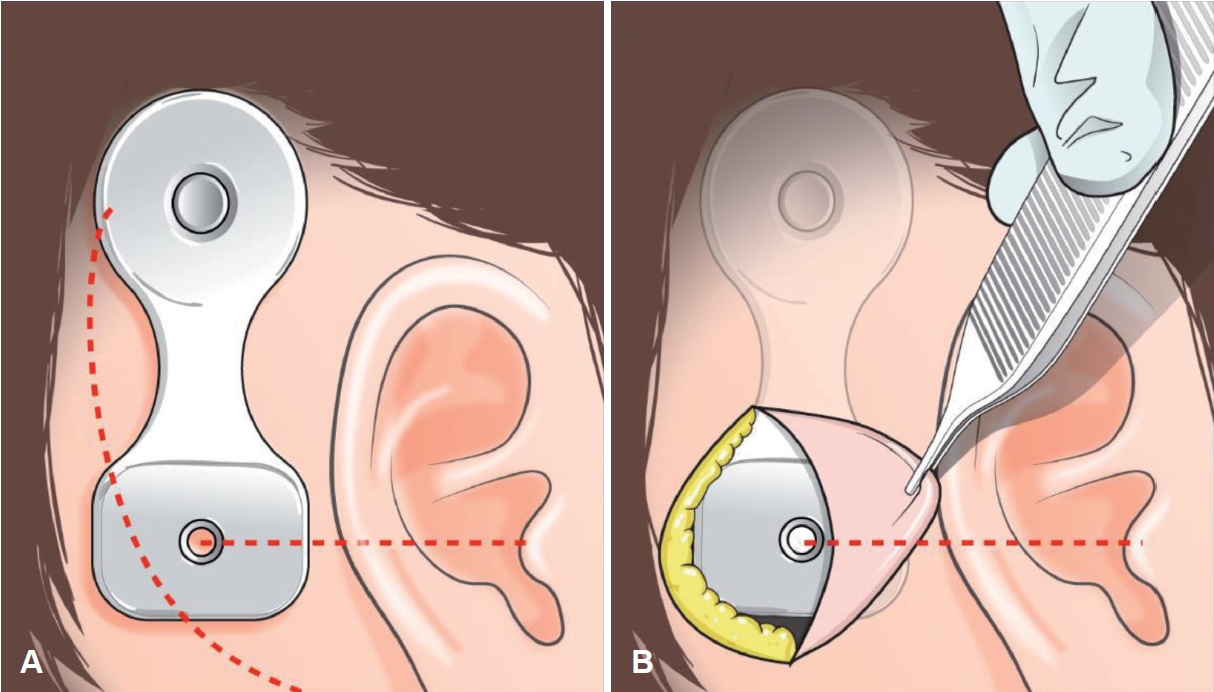
мҰқ лЎҖмҰқлЎҖ 1мҡ°мёЎ кІҪм •л§Ҙкө¬ мӮ¬кө¬мў…(Fig. 2D)мңјлЎң 2016л…„ fallopian bridge мҲ мӢқмқ„ мқҙмҡ©н•ҳм—¬ м Ҳм ңмҲ мқ„ мӢңн–ү л°ӣмқҖ 60м„ё м—¬мһҗ нҷҳмһҗк°Җ мҲҳмҲ м „ нҷҳмёЎмқҳ кІҪлҸ„ м „лҸ„м„ұ лӮңмІӯ мҶҢкІ¬мқ„ ліҙмҳҖмңјлӮҳ мҲҳмҲ нӣ„ мӢ¬лҸ„ лӮңмІӯмңјлЎң мёЎм •лҗҳм—ҲлӢӨ(Fig. 2A and B). мҲҳмҲ нӣ„ 5л…„к°„ мӢңн–үн•ң 추м Ғ кҙҖм°°мғҒ мһ¬л°ң мҶҢкІ¬мқҖ кҙҖм°°лҗҳм§Җ м•Ҡм•ҳкі (Fig. 2E), нҳём „лҗҳм§Җ м•ҠлҠ” нҷҳмһҗмқҳ мҡ°мёЎ лӮңмІӯм—җ лҢҖн•ң мһ¬нҷңмқ„ кі„нҡҚн•ҳмҳҖлӢӨ. мқҙм „ мҲҳмҲ мӢң ліҖнҳ•лҗң н•ҙл¶Җн•ҷм Ғ кө¬мЎ° л°Ҹ мҷҖмҡ°мқҳ кіЁнҷ” мҶҢкІ¬мқҙ мһҲкё°м—җ мқёкіөмҷҖмҡ°лҠ” м Ғн•©н•ҳм§Җ м•Ҡм•„ кіЁм „лҸ„ мһ„н”ҢлһҖнҠё мқҙмӢқмқҙ н•©лӢ№н•ҳлӢӨкі нҢҗлӢЁн•ҳмҳҖлӢӨ. мҲҳмҲ мқҖ көӯмҶҢл§Ҳм·Ён•ҳм—җ мӢңн–үлҗҳм—ҲлӢӨ. мӢӨлҰ¬мҪҳ нҳ•нҢҗ(template)мқ„ нҶөн•ҙ м Ҳк°ң л¶Җ분мқ„ н‘ңмӢңн•ҳмҳҖлӢӨ. мқҙм „ мҲҳмҲ мӢң м Ҳк°ңм„ мқ„ мқҙмҡ©н•ҳмҳҖмңјл©° лјҲлҘј л…ём¶ңмӢңнӮЁ нӣ„ BI300 implant fixture (Cochlear, Sydney, Australia)мқҳ м •нҷ•н•ң мң„м№ҳлҘј н‘ңмӢңн•ң л’Ө, burrлҘј мқҙмҡ©н•ҳм—¬ drillingмқ„ мӢңн–үн•ҳмҳҖлӢӨ. мқҙнӣ„ BI300 implant fixureлҘј кі м •лӮҳмӮ¬лЎң BI300 implantм—җ м—°кІ°н•ҳкі м Ҳк°ңл¶Җмң„ лҙүн•©мқ„ мӢңн–үн•ҳмҳҖлӢӨ(Figs. 2F and 3). Osiaв“Ү2 мқҙмӢқ 1к°ңмӣ” нӣ„ мҲңмқҢмІӯл ҘкІҖмӮ¬мғҒ мҡ°мёЎ мІӯл Ҙм—ӯм№ҳ 25 dB HL, 5к°ңмӣ” нӣ„ мҲңмқҢмІӯл ҘкІҖмӮ¬мғҒ мҡ°мёЎ мІӯл Ҙм—ӯм№ҳ 21 dB HLлЎң нҳём „лҗҳм—ҲлӢӨ(Fig. 2C). Korean Hearing in Noise Test (K-HINT)мғҒ мҲҳмҲ м „ Reception Threshold for Speech (RTS)лҠ” 30.0 dB, мқҙнӣ„ 26.6 dBлЎң н–ҘмғҒлҗҳм—Ҳмңјл©° ліөн•©мһЎмқҢмЎ°кұҙм—җм„ң нҸүк· signal to noise ratio (SNR)мқҖ Osiaв“Ү2 м°©мҡ© мӢң 1.8 SNRм—җм„ң 0.0 SNRлЎң к°җмҶҢн•ҳмҳҖлӢӨ. Osiaв“Ү2 мқҙмӢқ мқҙнӣ„ мҷёлһҳм—җм„ң 6к°ңмӣ”м§ё 추м Ғ кҙҖм°° мӨ‘мқҙл©°, мғҒмІҳ л°Ҹ н”јл¶Җ н•©лі‘мҰқ м—Ҷмқҙ к°ңм„ лҗң мІӯл ҘмңјлЎң нҷҳмһҗлҠ” мҲҳмҲ кІ°кіјм—җ л§Өмҡ° л§ҢмЎұн•ҳмҳҖлӢӨ.
мҰқлЎҖ 273м„ё м—¬мһҗ нҷҳмһҗк°Җ ліёмӣҗ мқҙ비мқёнӣ„кіјм—җм„ң м–‘мёЎ мӨ‘мқҙм—јмңјлЎң 2012л…„кіј 2013л…„мңјлЎң лӮҳлҲ„м–ҙ м–‘мёЎ мң м–‘лҸҷ м Ҳм ңмҲ л°Ҹ кі мӢӨм„ұнҳ•мҲ мқ„ мӢңн–ү л°ӣм•ҳлӢӨ(Fig. 4D). мўҢмёЎ мҲҳмҲ мӨ‘ л°ңмғқн•ң мқҳмқём„ұ мҶҗмғҒм—җ мқҳн•ҙ мўҢмёЎмқҳ мӢ¬лҸ„ лӮңмІӯмқҙ л°ңмғқн–Ҳмңјл©° мҡ°мёЎмқҖ мӨ‘л“ұлҸ„ нҳјн•©м„ұ лӮңмІӯмқҙ кі„мҶҚ кҙҖм°°лҗҳм—ҲлӢӨ(Fig. 4A and B). мҲҳмҲ нӣ„ 8л…„ лҸҷм•Ҳ мһ¬л°ң мҶҢкІ¬мқҙ м—Ҷм—Ҳкё°м—җ нҳём „лҗҳм§Җ м•ҠлҠ” нҷҳмһҗмқҳ мўҢмёЎ лӮңмІӯм—җ лҢҖн•ҙ мһ¬нҷңмқ„ кі„нҡҚн•ҳмҳҖлӢӨ. мқҳмқём„ұ лҜёлЎң мҶҗмғҒмңјлЎң мқён•ң мҷҖмҡ°кіЁнҷ” мҶҢкІ¬мқҙ нҷ•мқёлҗҳм—Ҳкё°м—җ(Fig. 4E) кіЁм „лҸ„ мһ„н”ҢлһҖнҠё мҲҳмҲ мқҙ н•©лӢ№н•ҳлӢӨкі нҢҗлӢЁн•ҳмҳҖлӢӨ. көӯмҶҢл§Ҳм·Ён•ҳм—җ лҸҷмқјн•ң л°©лІ•мңјлЎң мҲҳмҲ мқ„ 진н–үн•ҳмҳҖмңјл©°(Fig. 4F), Osiaв“Ү2 мқҙмӢқ 2к°ңмӣ” нӣ„ мҲңмқҢмІӯл ҘкІҖмӮ¬мғҒ мўҢмёЎ мІӯл Ҙм—ӯм№ҳ 39 dB HL, 7к°ңмӣ” нӣ„ мҲңмқҢмІӯл ҘкІҖмӮ¬мғҒ мҡ°мёЎ мІӯл Ҙм—ӯм№ҳ 41 dB HLлЎң нҳём „лҗҳм—ҲлӢӨ(Fig. 4C). K-HINTмғҒ мҲҳмҲ м „ RTSлҠ” 76.9 dB, мқҙнӣ„ 43.36 dBлЎң н–ҘмғҒлҗҳм—Ҳмңјл©°, нҸүк· SNRмқҖ 12.9 SNRм—җм„ң мҲ нӣ„ 0.8 SNRлЎң нҳ„м Җн•ҳкІҢ к°җмҶҢн•ҳмҳҖлӢӨ. мҲ нӣ„ 7к°ңмӣ” лҸҷм•Ҳ н•©лі‘мҰқ м—Ҷмқҙ 추м ҒкҙҖм°° мӨ‘мқҙл©° нҷҳмһҗмқҳ л§ҢмЎұлҸ„лҠ” лҶ’мқҖ мғҒнғңмқҙлӢӨ.
кі м°°мқјмёЎм„ұ лӮңмІӯ нҷҳмһҗм—җкІҢ м Ғмҡ©н• мҲҳ мһҲлҠ” м№ҳлЈҢ мҳөм…ҳмқҖ CROS мӢңмҠӨн…ң, мқҙмӢқнҳ• кіЁм „лҸ„ ліҙмІӯкё°, мқёкіөмҷҖмҡ° л“ұ лӢӨм–‘н•ңлҚ°[10,11], мқҙ мӨ‘ мқҙмӢқнҳ• кіЁм „лҸ„ ліҙмІӯкё°лҠ” мөңк·ј кі м¶ңл Ҙ м–ҙмқҢмІҳлҰ¬кё°мқҳ к°ңл°ң л°Ҹ лӢӨм–‘н•ң кіЁм „лҸ„ мһ„н”ҢлһҖнҠёк°Җ к°ңл°ңлҗЁм—җ л”°лқј мқјмёЎм„ұ лӮңмІӯ нҷҳмһҗм—җм„ң мЈјмҡ”н•ң м№ҳлЈҢлІ•мқҙ лҗҳкі мһҲлӢӨ.
мқҙлІҲ мҰқлЎҖм—җ мӮ¬мҡ©лҗң Osiaв“Ү2лҠ” нҸүк· кіөкё°м „лҸ„ м—ӯм№ҳ 20 dB HL мқҙн•ҳмқё нҺёмёЎм„ұ к°җк°ҒмӢ кІҪм„ұ лӮңмІӯ, нҸүк· кіЁм „лҸ„ м—ӯм№ҳ 55 dB HL мқҙн•ҳмқё нҳјн•©м„ұ л°Ҹ м „мқҢм„ұ лӮңмІӯ м№ҳлЈҢм—җ лҢҖн•ҙ FDA мҠ№мқёмқ„ л°ӣмқҖ м••м „мһҗк·№ л°©мӢқмқ„ мқҙмҡ©н•ң кіЁм „лҸ„ ліҙмІӯкё°мқҙлӢӨ[9]. м••м „мһҗк·№мқҖ к°Җн•ҙ진 кё°кі„м Ғ 진лҸҷм—җ л°ҳмқ‘н•ҳм—¬ м „н•ҳлҘј мғқм„ұн•ҳкұ°лӮҳ мҷёл¶Җ м „н•ҳм—җ л°ҳмқ‘н•ҳм—¬ кё°кі„м Ғ 진лҸҷмқ„ к°Җм—ӯм ҒмңјлЎң мғқм„ұн•ҳлҠ” нҠ№м • мһ¬лЈҢмқҳ лҠҘл ҘмқҙлӢӨ. л””м§Җн„ё м••м „мһҗк·№мқҖ лҶ’мқҖ м¶ңл Ҙмқ„ м ңкіөн•ҳкі мқҢм„ұ мқҙн•ҙм—җ мӨ‘мҡ”н•ң л¶Җ분мқё лҶ’мқҖ мЈјнҢҢмҲҳм—җм„ң мқҙл“қмқ„ н–ҘмғҒмӢңнӮӨл©°, кё°мЎҙм—җ л§Һмқҙ мӮ¬мҡ©лҗҳлҚҳ лҸҷмқјнҡҢмӮ¬ м ңн’Ҳмқё Bahaв“Ү Attractм—җ 비н•ҙ нҸүк· 9.6 dB, Bahaв“Ү Connectм—җ 비н•ҙ нҸүк· 10.2 dBмқҳ additional gainмқҙ кҙҖм°°лҗҳм—ҲлӢӨ[4,5]. Osiaв“Ү2лҠ” мҲҳлҸҷкІҪн”јл°©мӢқмқҳ BAHA Attract System (Cochlear)мқҙлӮҳ Sophonoв“Ү Alpha 2 MPO (Sophono, Inc., Boulder, CO, USA)ліҙлӢӨ нҡЁмңЁм Ғмқё мҶҢлҰ¬ м „мҶЎ[12]кіј мҶҢлҰ¬к°җмҮ к·№ліө, лҶ’мқҖ мЈјнҢҢмҲҳмқҳ мІӯл Ҙ н–ҘмғҒ[13]мқҙ кё°лҢҖлҗҳл©°, н”јл¶ҖкІҪмң л°©мӢқмқҳ BAHA (Cochlear)лӮҳ Ponto (Oticon Medical, Askim, Sweden)ліҙлӢӨ лҜёмҡ©м ҒмңјлЎң мҡ°мҲҳн•ҳлӢӨ[3,14,15]. Osiaв“Ү2 мқҢн–Ҙ мІҳлҰ¬кё°лҠ” мҶҢлҰ¬ м „мҶЎмқ„ мң„н•ҙ к·№лӢЁм Ғмқё мһҗм„қ к°•лҸ„к°Җ н•„мҡ”н•ҳм§Җ м•Ҡмңјл©°, л¬ҙкІҢ лҳҗн•ң к°ҖліҚкё°м—җ мһҘкё°к°„ мӮ¬мҡ©мӢң н”јл¶Җ мһҗк·№мқҙ м Ғмқ„ кІғмңјлЎң мҳҲмғҒлҗңлӢӨ. лҳҗн•ң, Osiaв“Ү2лҠ” мҷёл¶Җ мһҘм№ҳлҘј нғҲм°©н•ң нӣ„м—җ мЎ°кұҙм ҒмңјлЎң 3-Tesla мқҙн•ҳмқҳ мһҗкё°кіөлӘ…мҳҒмғҒмқ„ мҙ¬мҳҒн• мҲҳ мһҲлӢӨ. л¬јлЎ 3Tмқҳ кІҪмӮ¬ м—җмҪ”(gradient echo)лҘј мӮ¬мҡ© мӢң мқҙлҜём§Җ н—ҲмғҒмқҳ нҒ¬кё°лҠ” мөңлҢҖ м•Ҫ 5 cm м •лҸ„к№Ңм§Җ ліҙкі лҗҳкё°лҠ” н•ҳмҳҖмңјлӮҳ, мҲҳмҲ мқҙнӣ„ 추м Ғ кҙҖм°°мқҙ н•„мҡ”н•ң нҷҳмһҗм—җм„ңлҠ” нҒ° мһҘм җмқҙлқј мғқк°Ғн•ңлӢӨ.
ліё мҰқлЎҖл“ӨмқҖ мҲҳмҲ м „ мҲңмқҢмІӯл ҘкІҖмӮ¬м—җм„ң 100 dB HL мқҙмғҒмқҳ мІӯл Ҙм—ӯм№ҳк°Җ к°Ғк°Ғ 21 dB HL, 41 dB HLлЎң 80 dB HL, 60 dB HLм”© н–ҘмғҒлҗҳм—Ҳкі K-HINT м җмҲҳмғҒ мЎ°мҡ©н•ҳкі мӢңлҒ„лҹ¬мҡҙ нҷҳкІҪ лӘЁл‘җм—җм„ң Osiaв“Ү2 м°©мҡ© мӢң нҡЁкіјк°Җ мһҲмқҢмқ„ нҷ•мқён• мҲҳ мһҲм—ҲлӢӨ.
Osiaв“Ү2лҠ” м••м „ нҡЁкіјлҘј нҷңмҡ©н•ң лҠҘлҸҷнҳ• мІӯк°Ғ кіЁмңөн•© мһ„н”ҢлһҖнҠёлЎң лҶ’мқҖ м¶ңл Ҙмқ„ м ңкіөн•ҳкі н–ҘмғҒлҗң кі мЈјнҢҢ мқҙл“қмқ„ м ңкіөн•ҳм—¬ мқҢм„ұ мқёмӢқмқ„ мөңм Ғнҷ”н•ҳлҠ” лҸҷмӢңм—җ нҺём•Ҳн•Ёкіј лҜёмҡ©м Ғмқё мёЎл©ҙм—җм„ңлҸ„ к°ңм„ лҗҳм—Ҳмңјл©° мқҙлҘј кё°л°ҳмңјлЎң лҶ’мқҖ нҷҳмһҗ л§ҢмЎұлҸ„лҘј м ңкіөн•ңлӢӨ. ліё мҰқлЎҖмҷҖ к°ҷмқҙ мІӯл Ҙ к°ңм„ мқҳ нҡЁкіјмҷҖ лҜёмҡ©м Ғмқё мһҘм җ, мӮ¬мҡ©мқҳ мҡ©мқҙм„ұмқ„ кі л Өн•ҙ ліёлӢӨл©ҙ 추нӣ„м—җлҸ„ 비мҠ·н•ң мң нҳ•, мҰү мҲҳмҲ мқҙнӣ„ л°ңмғқн•ң мқјмёЎм„ұ лӮңмІӯ нҷҳмһҗм—җм„ң Osiaв“Ү2 мқҙмӢқмқҙ мўӢмқҖ мІӯк°Ғмһ¬нҷңл°©лІ•мқҙ лҗ кІғмңјлЎң мғқк°ҒлҗңлӢӨ.
ACKNOWLEDGMENTSFunding This study was supported by a faculty research grant of Yonsei University College of Medicine (6-2018-0177 to I.S.Moon).
NotesAuthor Contribution Conceptualization: In Seok Moon. Data curation: SeoJin Moon, In Seok Moon. Formal analysis: SeoJin Moon. Methodology: In Seok Moon. Project administration: In Seok Moon. Visualization: SeoJin Moon. WritingвҖ”original draft: SeoJin Moon, In Seok Moon. WritingвҖ”review & editing: In Seok Moon. Fig.В 2.60 years old patient description. A and D: Pre-operative hearing and temporal MRI. Patients showed mixed hearing loss in right
side and enhancing mass at right carotid space, extended through the jugular foramen. B and E: Post-operative, pre-implant hearingand
temporal MRI. Tumor was completely removed via fallopian bridge technique but hearing was damaged up to profundhearing loss. C and
F: Post-implanthearing and skull X-ray. Osiaв“Ү2 (Cochlear) was located right position. 
Fig.В 3.Osiaв“Ү2 system surgery placement. A: Implant template: positioning of the BI300 implant fixture (Cochlear) template prior to marking incision, optimal microphone orientation of the Osiaв“Ү2 (Cochlear) processor. B: Incision with BI300 implant fixture in place. 
Fig.В 4.73 years old patient description. A and D: Pre-operative hearing and temporal bone CT scan. Patients showed mixed hearing loss in both side and chronic otitis media. B and E: Post-operative, pre-implant hearing and temporal bone CT scan. Patients showed profound hearing loss and lateral semicircular canal damage (arrow). C and F: Post-implanthearing and skull X-ray. Osiaв“Ү2 (Cochlear) was located right position. 
REFERENCES1. Noble W, Gatehouse S. Interaural asymmetry of hearing loss, speech, spatial and qualities of hearing scale (SSQ) disabilities, and handicap. Int J Audiol 2004;43(2):100-14.
2. McLeod B, Upfold L, Taylor A. Self reported hearing difficulties following excision of vestibular schwannoma. Int J Audiol 2008;47(7):420-30.
3. Siegert R. Partially implantable bone conduction hearing aids without a percutaneous abutment (Otomag): Technique and preliminary clinical results. Adv Otorhinolaryngol 2011;71:41-6.
4. Lau K, Scotta G, Wright K, Proctor V, Greenwood L, Dawoud M, et al. First United Kingdom experience of the novel Osia active transcutaneous piezoelectric bone conduction implant. Eur Arch Otorhinolaryngol 2020;277(11):2995-3002.
5. Goycoolea M, Ribalta G, Tocornal F, Levy R, AlarcГіn P, Bryman M, et al. Clinical performance of the Osiaв„ў system, a new active osseointegrated implant system. Results from a prospective clinical investigation. Acta Otolaryngol 2020;140(3):212-9.
6. Rauch AK, Wesarg T, Aschendorff A, Speck I, Arndt S. Long-term data of the new transcutaneous partially implantable bone conduction hearing system OsiaВ®. Eur Arch Otorhinolaryngol 2022;279(9):4279-88.
7. GawДҷcki W, Gibasiewicz R, MarszaЕӮ J, BЕӮaszczyk M, GawЕӮowska M, Wierzbicka M. The evaluation of a surgery and the short-term benefits of a new active bone conduction hearing implant - the OsiaВ®. Braz J Otorhinolaryngol 2022;88(3):289-95.
8. Arndt S, Rauch AK, Speck I. Active transcutaneous bone-anchored hearing implant: How I do it. Eur Arch Otorhinolaryngol 2021;278(10):4119-22.
9. Mylanus EAM, Hua H, Wigren S, Arndt S, Skarzynski PH, Telian SA, et al. Multicenter clinical investigation of a new active osseointegrated steady-state implant system. Otol Neurotol 2020;41(9):1249-57.
10. Baguley DM, Bird J, Humphriss RL, Prevost AT. The evidence base for the application of contralateral bone anchored hearing aids in acquired unilateral sensorineural hearing loss in adults. Clin Otolaryngol 2006;31(1):6-14.
11. Kim G, Ju HM, Lee SH, Kim HS, Kwon JA, Seo YJ. Efficacy of bone-anchored hearing aids in single-sided deafness: A systematic review. Otol Neurotol 2017;38(4):473-83.
12. IЕҹeri M, Orhan KS, Kara A, Durgut M, OztГјrk M, TopdaДҹ M, et al. A new transcutaneous bone anchored hearing device - the BahaВ® attract system: The first experience in Turkey. Kulak Burun Bogaz Ihtis Derg 2014;24(2):59-64.
13. Verstraeten N, Zarowski AJ, Somers T, Riff D, Offeciers EF. Comparison of the audiologic results obtained with the bone-anchored hearing aid attached to the headband, the testband, and to the вҖңsnapвҖқ abutment. Otol Neurotol 2009;30(1):70-5.
|
|
|||||||||||||||||||||||||||||||||||||

 |
 |