 |
 |
AbstractNasal dermoids are congenital midline nasal lesions that occur along with encephaloceles and gliomas. They can cause both deformity of nasal structure and intracranial infection as they grow. Treatment for these lesions is be concerned with two aspects, the complete removal of the lesions and making the surgical scar cosmetically acceptable. To that goal, many surgical approaches such as vertical incision, transverse incision, lateral rhinotomy and open rhinoplasty have been introduced. A 12-month male child presented with palpable mass at nasal root. The mass was easily movable, non-compressible and did not present fistula. A well-defined cystic mass without intracranial extension was found on the computerized tomography scans. Open rhinoplasty approach was opted for according to the guardiansŌĆÖ preference to avoid visible facial scar, and the lesions were completely resected. The pathologic examination confirmed the lesion to be nasal dermoids. The columellar scar was negligible and there was no recurrence at 5 year-follow up after surgery.
ņä£ ļĪĀņĮöņŚÉ ļ░£ņāØĒĢśļŖö ņäĀņ▓£ņä▒ ņĀĢņżæņäĀ ņóģņ¢æņØĆ ļō£ļ¼Ė ļ│æļ│Ćņ£╝ļĪ£ ļćīļźś, ņŗĀĻ▓ĮĻĄÉņóģ, ļ╣äņ£ĀĒö╝ņóģņØä ĒżĒĢ©ĒĢśņŚ¼ ņØĖĻĄ¼ 10ļ¦ī ļ¬ģļŗ╣ 2.5ļ¬ģņŚÉņä£ 5ļ¬ģ Ļ╝┤ļĪ£ ļ░£ņāØĒĢ£ļŗż[1]. ļ╣äņ£ĀĒö╝ņóģņØĆ ļéŁ, ļÅÖ, ļłäĻ│ĄņØś ĒśĢĒā£ļĪ£ ņĪ┤ņ×¼ĒĢśļ®░, ļō£ļ¼╝Ļ▓ī ļæÉĻ░£ļé┤ Ļ│ĄĻ░ä(intracranial space)Ļ╣īņ¦Ć ņ╣©ļ▓öĒĢśĻĖ░ļÅä ĒĢ£ļŗż[2]. ļ│æļ│ĆņØś Ēü¼ĻĖ░ņÖĆ ņ£äņ╣ś, ņ╣©ļ▓ö ņĀĢļÅäļź╝ ĒÅēĻ░ĆĒĢśĻĖ░ ņ£äĒĢ┤ ņĀäņé░ĒÖöļŗ©ņĖĄņ┤¼ņśü(CT) ļ░Å ņ×ÉĻĖ░Ļ│Ąļ¬ģņśüņāü(MRI) ņ┤¼ņśüņØ┤ ĒĢäņÜöĒĢśļŗż[3]. ļ│æļ│Ćņ£╝ļĪ£ ņØĖĒĢ£ ņŻ╝ļ│Ć ņĪ░ņ¦üņØś ļ│ĆĒśĢ ļ░Å ĻĄŁņåī Ļ░ÉņŚ╝, ļćīļ¦ēņŚ╝ ļśÉļŖö ļćīļåŹņ¢æņŚÉ ļīĆĒĢ£ ĒĢ®ļ│æņ”Ø Ļ░ĆļŖźņä▒ ļĢīļ¼ĖņŚÉ ņĀüņĀłĒĢ£ ņŗ£ĻĖ░ņŚÉ ņÖäņĀäĒĢ£ ņłśņłĀņĀü ņĀłņĀ£Ļ░Ć ĒĢäņÜöĒĢśĻ│Ā, ņłśņłĀ ņŗ£ĻĖ░ņÖĆ ļ░®ļ▓ĢņØĆ ņ”ØļĪĆņŚÉ ļö░ļØ╝ Ļ░£ļ│äņĀüņ£╝ļĪ£ Ļ│ĀļĀżĒĢ┤ņĢ╝ ĒĢ£ļŗż[2].
ņĀĆņ×ÉļōżņØĆ ļ╣äņ£ĀĒö╝ņóģņ£╝ļĪ£ ņ¦äļŗ©ļ░øņØĆ 12Ļ░£ņøö ļé©ņĢäņŚÉ ļīĆĒĢ┤ Ļ░£ļ░® ņĮöņä▒ĒśĢ ņĀæĻĘ╝ļ▓Ģ(open rhinoplasty approach)ņ£╝ļĪ£ ņÖäņĀä ņĀłņĀ£ Ēøä 5ļģäņ¦ĖĻ╣īņ¦Ć ĒŖ╣ļ│äĒĢ£ ĒĢ®ļ│æņ”Ø ļ░Å ņ×¼ļ░£ ņŚåņØ┤ Ļ▓ĮĻ│╝Ļ┤Ćņ░░ ņżæņ£╝ļĪ£ ņØ┤ļź╝ ļ¼ĖĒŚīĻ│Āņ░░Ļ│╝ ĒĢ©Ļ╗ś ļ│┤Ļ│ĀĒĢśĻ│Āņ×É ĒĢ£ļŗż.
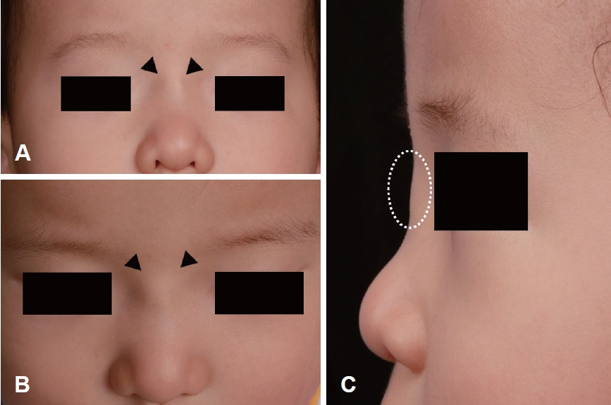
ņ”Ø ļĪĆ12Ļ░£ņøö ļé©ņĢäĻ░Ć ļ╣äļ░░ļČĆņŚÉ ļ¦īņĀĖņ¦ĆļŖö ņóģļ¼╝ņØä ņŻ╝ņåīļĪ£ ļé┤ņøÉĒĢśņśĆļŗż. ņČ£ņāØ ļ░Å ņŗĀņāØņĢäĻĖ░ ĒŖ╣ļ│äĒĢ£ ļ¼ĖņĀ£ļŖö ņŚåņŚłĻ│Ā, Ļ░ĆņĪ▒ļĀźņŚÉņä£ ĒŖ╣ņØ┤ņé¼ĒĢŁņØĆ ņŚåņŚłļŗż. ņŗĀņ▓┤ ņ¦äņ░░ņŗ£ ņóģļ¼╝ņØĆ ļ╣äļ░░ļČĆņŚÉ ņ£äņ╣śĒĢ£ 1 cm Ēü¼ĻĖ░ņØś ļ╣äĻĄÉņĀü ļŗ©ļŗ©ĒĢśĻ▓ī ņ┤ēņ¦ĆļÉśļŖö ņøÉĒśĢņØś Ēö╝ĒĢś ņóģļ¼╝ļĪ£ ņĢĢĒåĄņØĆ ņŚåņŚłņ£╝ļ®░ ļ¦īņĪīņØä ļĢī Ļ│ĀņĀĢļÉśņ¢┤ ņ׳ņ¦Ć ņĢŖĻ│Ā ņēĮĻ▓ī ņøĆņ¦üņØ┤ļŖö ņ¢æņāüņØ┤ņŚłļŗż(Fig. 1). ņŗĀņ▓┤ņ¦äņ░░ņŚÉņä£ ĻĘĖ ļ░¢ņØś ņØ┤ņāü ņåīĻ▓¼ņØĆ Ļ┤Ćņ░░ļÉśņ¦Ć ņĢŖņĢśļŗż.
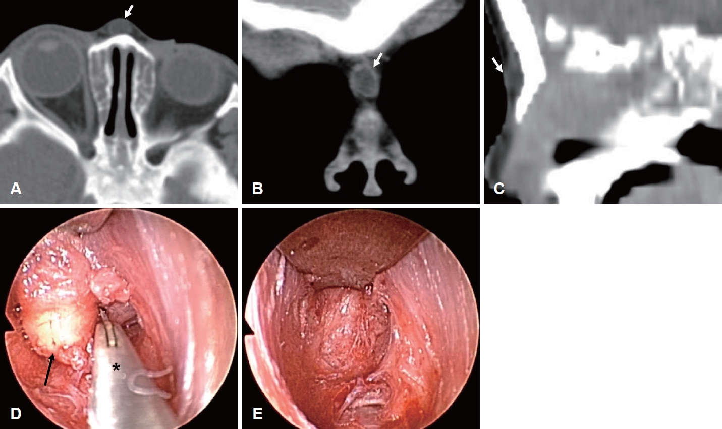
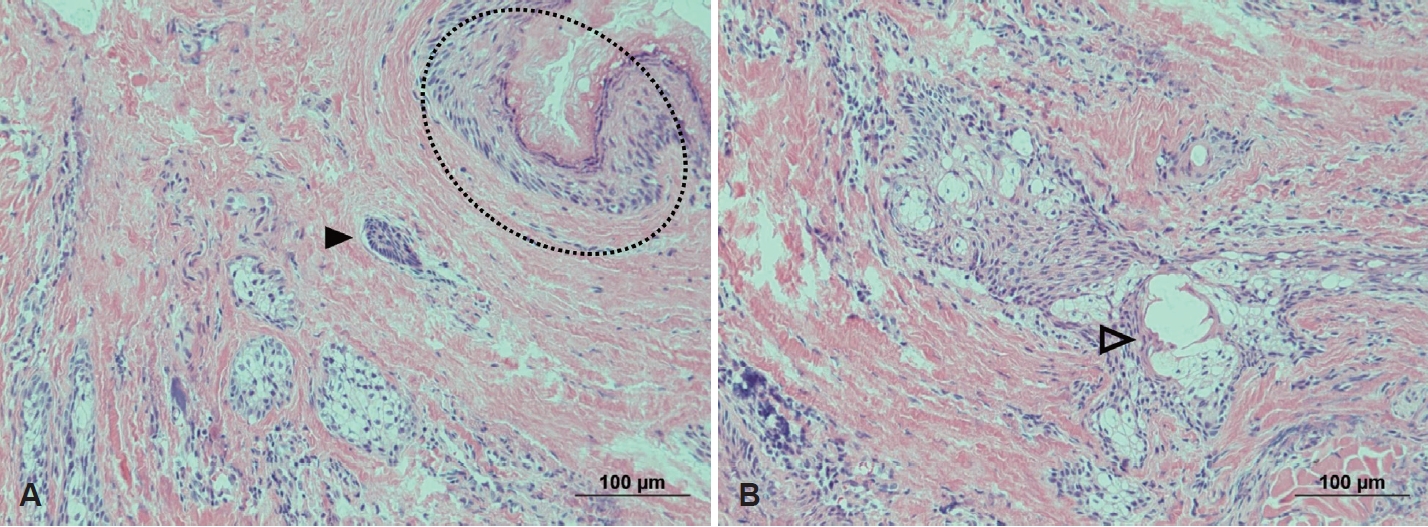
ļ│æļ│ĆņØś Ēü¼ĻĖ░, ļ¬©ņ¢æ ļ░Å ļ▓öņ£äļź╝ ĒÅēĻ░ĆĒĢśĻĖ░ ņ£äĒĢ┤ ņ┤¼ņśüĒĢ£ ņĪ░ņśüņ”ØĻ░Ģ CTņŚÉņä£ ņóģļ¼╝ņØĆ 0.8 cm Ēü¼ĻĖ░ņØś ļ╣äĻĘ╝ļČĆ Ēö╝ĒĢśņ¦Ćļ░®ņĖĄņŚÉ ĻĄŁĒĢ£ļÉ£ ļéŁņä▒ ļ│æļ│Ćņ£╝ļĪ£ ĒÖĢņØĖļÉśņŚłĻ│Ā, ļæÉĻ░£ļé┤ ņ╣©ļ▓ö ņåīĻ▓¼ņØĆ ņŚåņŚłļŗż(Fig. 2). ņĀäņŗĀļ¦łņĘ©ĒĢś Ļ░£ļ░®ņĮöņä▒ĒśĢņĀæĻĘ╝ļ▓Ģņ£╝ļĪ£ ņóģļ¼╝ ņĀłņĀ£ļź╝ ņŗ£Ē¢ēĒĢśņśĆļŗż. ļ╣äņŻ╝ņØś ņŚŁ VĒśĢ ņĀłĻ░£(inverted V shape incision) ļ░Å ņ¢æņĖĪ ļ╣äņØĄņØś ļ│ĆņŚ░ ņĀłĻ░£(marginal incision) Ēøä ņŚ░Ļ│©ļ¦ē ļ░Å Ļ│©ļ¦ēņ£ä ņĀłĻ░£ļ®┤(supraperichondrial and supraperiosteal plane)ņ£╝ļĪ£ ļ░Ģļ”¼ļź╝ ņŗ£Ē¢ēĒĢśņśĆļŗż. 4 mm 0┬░ Ļ░Ģņ¦üĒśĢ ļé┤ņŗ£Ļ▓Į ņŗ£ņĢ╝ĒĢśņŚÉ ļ╣äļ░░ļČĆĻ╣īņ¦Ć ļ░Ģļ”¼ĒĢśņŚ¼ ļ╣äĻĘ╝ļČĆņŚÉ ĻĄŁĒĢ£ļÉ£ ļéŁņóģņØä ĒÖĢņØĖĒĢśĻ│Ā ļéŁņóģņØ┤ ĒīīņŚ┤ļÉśņ¦Ć ņĢŖļÅäļĪØ ņŻ╝ļ│Ć ņĪ░ņ¦üĻ│╝ņØś ļ░Ģļ”¼ļź╝ ņŗ£Ē¢ēĒĢśņśĆļŗż. ĒĢśņ¦Ćļ¦ī, ļé┤ņŗ£Ļ▓Į ņŗ£ņĢ╝ ļÆĘĒÄĖņØĖ ļæÉņĖĪ(cephalic)ņØś ņŻ╝ļ│Ć ņĪ░ņ¦üĻ│╝ ļ░Ģļ”¼ļź╝ ņ£äĒĢ┤ ļéŁņóģņØä Ļ▓¼ņØĖĒĢśļŗż ļéŁņóģ ņØ╝ļČĆĻ░Ć ĒīīņŚ┤ņØ┤ ļÉśņ¢┤ ļéŁņóģļé┤ ņ╝ĆļØ╝Ēŗ┤ ļ¼╝ņ¦łļōżņØ┤ ņØ╝ļČĆ ņ£ĀņČ£ļÉśņ¢┤ ļéŁņóģ ņÖäņĀä ņĀłņĀ£ Ēøä ļŗżļ¤ēņØś ņāØļ”¼ņŗØņŚ╝ņłś ņäĖņ▓ÖņØä ņŚ¼ļ¤¼ ņ░©ļĪĆ ņŗ£Ē¢ēĒĢśņśĆļŗż. ņłśņłĀ Ēøä ļ╣äĻ░Ģ Ēī®Ēé╣ņØĆ ņŗ£Ē¢ēĒĢśņ¦Ć ņĢŖņĢśĻ│Ā, ņÖĖļ╣äņØś ļČĆņóģ ļ░Å ņןņĢĪņóģ ļ░£ņāØņØä ņśłļ░®ĒĢśĻĖ░ ņ£äĒĢ┤ ņŚ┤Ļ░Ćņåīņä▒ ļČĆļ¬®(thermoplastic aqua splint)ņØä ņÖĖļ╣äņŚÉ Ļ▒░ņ╣śĒĢśņśĆņ£╝ļ®░ ņłśņłĀ Ēøä 3ņØ╝ņ¦Ė Ļ▓Įļ»ĖĒĢ£ ļČĆņóģĻ│╝ ļ░śņāüņČ£Ēśł(ecchymosis)ļ¦ī ļ│┤ņŚ¼ ņĀ£Ļ▒░ĒĢśņśĆļŗż. ņĀłņĀ£ĒĢ£ ņóģļ¼╝ņØś ļ│æļ”¼ņĪ░ņ¦üĻ▓Ćņé¼ņŚÉņä£ ņ╝ĆļØ╝Ēŗ┤ĒÖö ļÉśņ¢┤ ņ׳ļŖö ņżæņĖĄ ĒÄĖĒÅē ņāüĒö╝ņÖĆ ļ¬©ļéŁ, Ēö╝ņ¦Ćņāś ļō▒ņØś ņåīĻ▓¼ņØ┤ ļ│┤ņŚ¼ ļ╣äņ£ĀĒö╝ņóģņ£╝ļĪ£ ĒÖĢņ¦äļÉśņŚłļŗż(Fig. 3). ņłĀĒøä ĒĢ£ļŗ¼ ņ¦Ė ņÖĖļלņŚÉņä£ ļ╣äņŻ╝ņØś ņĀłĻ░£ ļČĆņ£ä ĒØēĒä░ ņÖĖņŚÉ ĒŖ╣ņØ┤ņåīĻ▓¼ņØĆ ņŚåņŚłņ£╝ļ®░(Fig. 4), ņØ┤Ēøä ļ│┤ĒśĖņ×ÉĻ░Ć ņÖĖļל ļ░®ļ¼ĖņŚÉ ņØæĒĢśņ¦Ć ņĢŖņĢä ņĀäĒÖö ņĪ░ņé¼ļź╝ ĒåĄĒĢ┤ ĒÖĢņØĖĒĢśņśĆņØä ļĢī ņłĀĒøä 5ļģäĻ╣īņ¦Ć ņóģļ¼╝ņØś ņ×¼ļ░£ņØĆ ņŚåņŚłĻ│Ā, ļłłņŚÉ ļØäļŖö ļ╣äņŻ╝ņØś ņĀłĻ░£ ļ░śĒØöļÅä ņŚåņŚłļŗżĻ│Ā ĒĢśņśĆļŗż.
Ļ│Ā ņ░░ļ╣äņ£ĀĒö╝ņóģņØĆ ņĮöņØś ņĀĢņżæņäĀņŚÉņä£ ļ░£ņāØĒĢĀ ņłś ņ׳ļŖö ņäĀņ▓£ņä▒ ļéŁņóģņ£╝ļĪ£ ļćīļźś, ņŗĀĻ▓ĮĻĄÉņóģĻ│╝ Ļ░Éļ│äņØ┤ ĒĢäņÜöĒĢśļŗż[1,2]. Ēā£ņāØĻĖ░ņŚÉ ņĀäļ╣äĻ░ĢĻ│╝ ņĀäļæÉņ▓£ļ¼ĖņŚÉņä£ ņĪ┤ņ×¼ĒĢśļŖö ņŗĀĻ▓ĮņÖĖļ░░ņŚĮ ņĪ░ņ¦üļōżņØĆ ņĀÉņ░© ļ│ĄĻĘĆļÉśņ¢┤ ņé¼ļØ╝ņ¦ĆļŖöļŹ░, ņØ╝ļČĆ Ēć┤Ē¢ēņŚÉ ņŗżĒī©ĒĢśĻ│Ā ņĀĢņżæņäĀņØä ļö░ļØ╝ ļéŁ ĒśĢĒā£ļĪ£ ņ×öņĪ┤ņŗ£ ļ╣äņ£ĀĒö╝ņóģņØ┤ ĒśĢņä▒ļÉĀ ņłś ņ׳ļŗżļŖö ņĀäļ╣äĻ│ĄĻ░ä(prenasal space) ņØ┤ļĪĀņØ┤ ļ░£ņāØĻĖ░ņĀäņ£╝ļĪ£ ņäżļ¬ģļÉśĻ│Ā ņ׳ļŗż[3,4]. ļ╣äņ£ĀĒö╝ņóģņØĆ ļ╣äņŻ╝ņŚÉņä£ ļ╣äļ░░ļČĆļź╝ ļö░ļØ╝ ļ»ĖĻ░ä ņé¼ņØ┤Ļ╣īņ¦Ć ņóģļ¼╝, ļłäĻ│Ą ļ░Å ļÅÖļ░śļÉ£ ļåŹņä▒ ļČäļ╣äļ¼╝ ļō▒ņ£╝ļĪ£ ļ░£Ļ▓¼ļÉ£ļŗż[5]. ļ╣äņ£ĀĒö╝ņóģņØĆ ļ╣äĻĄÉņĀü ļŗ©ļŗ©ĒĢśĻ│Ā ļ╣äņĢĢņČĢņä▒ņ£╝ļĪ£ ļ╣øņØ┤ ņל Ēł¼Ļ│╝ļÉśņ¦Ć ņĢŖļŖöļŗż[2]. ņÜĖņŚłņØä ļĢī ļ│æļ│ĆņØ┤ ņ╗żņ¦Ćņ¦Ć ņĢŖĻ│Ā, Ļ▓ĮņĀĢļ¦źņØä ņĢĢļ░ĢĒĢśņśĆņØä ļĢī Ēü¼ĻĖ░Ļ░Ć ņ╗żņ¦ĆļŖöņ¦Ć ĒÖĢņØĖĒĢśļŖö Furstenberg testņŚÉņä£ ņØīņä▒ņØä ļ│┤ņØĖļŗż[2,3]. ņØ┤ņÖĆ ļŗ¼ļ”¼ ļćīļźśļŖö ļČĆļō£ļ¤ĮĻ│Ā, ņĢĢļ░Ģņŗ£ ņל ļłīļĀżņ¦Ćļ®░, ļ░ĢļÅÖņä▒ņØ┤ ņ׳Ļ│Ā, ļ╣øņŚÉ ņל Ēł¼Ļ│╝ļÉśļ®░, Furstenberg test ņ¢æņä▒ ņåīĻ▓¼ņØä ļ│┤ņØĖļŗż[6]. ļśÉ ļŗżļźĖ Ļ░Éļ│äņ¦äļŗ© ļ│æļ│ĆņØĖ ņŗĀĻ▓ĮĻĄÉņóģņØĆ ļŗ©ļŗ©ĒĢśĻ▓ī ļ¦īņĀĖņ¦Ćļ®░ ļ¬©ņäĖĒśłĻ┤ĆņØś ĒÖĢņןņØ┤ ļ│┤ņØ╝ ņłś ņ׳Ļ│Ā, Furstenberg test ņØīņä▒ņØä ļ│┤ņØ┤ļ®░ ņłśļ¦ēņ£╝ļĪ£ ņŚ░Ļ▓░ļÉśņ¦Ć ņĢŖļŖöļŗż[3,6]. ņĀĢĒÖĢĒĢ£ ņ¦äļŗ© ļ░Å ļæÉĻ░£ļé┤ ņ╣©ļ▓ö ņĀĢļÅäļź╝ ĒÖĢņØĖĒĢśĻĖ░ ņ£äĒĢ┤ņäĀ ņśüņāü Ļ▓Ćņé¼Ļ░Ć ĒĢäņłśņĀüņØ┤ļ®░ CTņÖĆ MRIļź╝ ņŻ╝ļĪ£ ņØ┤ņÜ®ĒĢśĻ▓ī ļÉ£ļŗż[5,7]. CTņāü ļæÉĻ░£ļé┤ ņ╣©ļ▓ö ņåīĻ▓¼ņØ┤ ļ¬ģĒÖĢĒĢśņ¦Ć ņĢŖņØĆ Ļ▓ĮņÜ░ MRI Ļ▓Ćņé¼Ļ░Ć ņČöĻ░ĆņĀüņ£╝ļĪ£ ĒĢäņÜöĒĢśļŗż[8]. CTņŚÉņä£ ņØ┤ļČäĒÖöļÉ£ Ļ│äĻ┤ĆņØ┤ ļ│┤ņØ┤Ļ▒░ļéś, Ļ│äĻ┤ĆņØś Ļ│© Ļ▓░ņåÉ ļśÉļŖö ļ¦╣Ļ│ĄņØś Ēü¼ĻĖ░Ļ░Ć ņ”ØĻ░ĆļÉ£ ņåīĻ▓¼ ļō▒ņØ┤ ļ│┤ņØ╝ Ļ▓ĮņÜ░, ļæÉĻ░£ļé┤ ņ╣©ļ▓ö Ļ░ĆļŖźņä▒ņØä ņØśņŗ¼ĒĢ┤ ļ│╝ ņłś ņ׳ļŗż[7]. ĒĢśņ¦Ćļ¦ī ņåīņĢäņØś Ļ▓ĮņÜ░, ņé¼Ļ│©ņØś ņłśņ¦üĒīÉĻ│╝ Ļ│äĻ┤ĆņØś ļČłņÖäņĀäĒĢ£ Ļ│©ĒÖöļĪ£ ļæÉĻ░£ļé┤ ņ╣©ļ▓ö ņ£äņ¢æņä▒ ņåīĻ▓¼ņØä ļéśĒāĆļé┤ņ¢┤ Ļ░Éļ│äņØä ņ¢┤ļĀĄĻ▓ī ĒĢĀ ņłś ņ׳ļŗż[9]. Bloom ļō▒[9]ņØĆ ņóģļ¼╝ņØ┤ ļ»ĖĻ░äņŚÉ ņĪ┤ņ×¼ĒĢśņ¦Ć ņĢŖĻ│Ā, ļ╣äĻ│©ļ│┤ļŗż Ēæ£ļ®┤ņŚÉ ņ׳Ļ│Ā, ņĀĢņāü Ļ│äĻ┤ĆņØś ĒśĢĒā£Ļ░Ć ļ│┤ņØ╝ ļĢī, CTļ¦īņ£╝ļĪ£ļÅä ļæÉĻ░£ļé┤ ņ╣©ļ▓ö ņŚ¼ļČĆļź╝ ļ░░ņĀ£ĒĢĀ ņłś ņ׳ļŗżĻ│Ā ĒĢśņśĆļŗż. ņāüĻĖ░ ĒÖśņĢäņØś Ļ▓ĮņÜ░ CTņŚÉņä£ ņóģļ¼╝ņØ┤ ļ╣äĻĘ╝ļČĆņŚÉ ļ╣äĻ│©ļ│┤ļŗż Ēæ£ļ®┤ņŚÉ ņĪ┤ņ×¼ĒĢśĻ│Ā, ļæÉĻ░£ļé┤ ņ╣©ļ▓ö ņåīĻ▓¼ņØ┤ ņŚåņŚłĻĖ░ ļĢīļ¼ĖņŚÉ ņČöĻ░ĆņĀüņØĖ ņśüņāü Ļ▓Ćņé¼ļŖö ņŗ£Ē¢ēĒĢśņ¦Ć ņĢŖņĢśļŗż. ņĀ£Ļ▒░ ņŗ£ĻĖ░ņŚÉ ļīĆĒĢ£ ņØśĻ▓¼ņØĆ ļŗżņ¢æĒĢśļéś, ņóģļ¼╝ņØś Ēü¼ĻĖ░Ļ░Ć ņ╗żņ¦Ćļ®┤ņä£ ļ░£ņāØĒĢĀ ņłś ņ׳ļŖö ņŻ╝ļ│Ć ņĪ░ņ¦üņØś ļ│ĆĒśĢ, ĻĄŁņåī Ļ░ÉņŚ╝ņØ┤ļéś ļæÉĻ░£ļé┤ ņ╣©ļ▓öņ£╝ļĪ£ ņØĖĒĢ£ ĒĢ®ļ│æņ”Ø Ļ░ĆļŖźņä▒ņØ┤ ņ׳ņ£╝ļ»ĆļĪ£ Ļ░ĆļŖźĒĢ£ ļ╣©ļ”¼ ņĀ£Ļ▒░ĒĢśļŖö Ļ▓āņØ┤ ļ░öļ×īņ¦üĒĢśļŗż[7]. Ļ│╝Ļ▒░ ĻĄŁļé┤ņŚÉņä£ ļ│┤Ļ│ĀļÉśņŚłļŹś ļ╣äņ£ĀĒö╝ņóģņØś Ļ▓ĮņÜ░ņŚÉļÅä 5ņäĖņŚÉ ņØ┤ļ»Ė ļ╣äĻ│©ņØś Ļ│©ĒīīĻ┤┤Ļ░Ć ļÅÖļ░śļÉśņŚłņØīņØä ĒÖĢņØĖĒĢĀ ņłś ņ׳ļŗż[10]. ņĀĆņ×ÉļōżņØś Ļ▓ĮņÜ░ņŚÉļÅä ĒÖśņĢäņØś ļéśņØ┤Ļ░Ć ņ¢┤ļ”░ ņĀÉņØä Ļ░ÉņĢłĒĢśņŚ¼ Ļ▓ĮĻ│╝ Ļ┤Ćņ░░ĒĢśļ®░ ņĀüņĀłĒĢ£ ņłśņłĀ ņŗ£ĻĖ░ļź╝ Ļ│Āļ»╝ĒĢśņśĆņ£╝ļéś, Ļ▓ĮĻ│╝ Ļ┤Ćņ░░ņŗ£ ņóģļ¼╝ņØ┤ ņ×ÉļØ╝ļ®┤ņä£ ļ╣äĻ│© ļ░Å ļ╣äņŚ░Ļ│© ļ│ĆĒśĢņ£╝ļĪ£ ņØĖĒĢ┤ 2ņ░©ņĀüņØĖ ņÖĖļ╣äņä▒ĒśĢņłĀņØ┤ ĒĢäņÜöĒĢśĻ▓ī ļÉĀ ņłś ņ׳ņ¢┤ ļ░öļĪ£ ņłśņłĀņØä ņ¦äĒ¢ēĒĢśĻĖ░ļĪ£ ĒĢśņśĆļŗż. ņłśņłĀ ņŗ£ ļČłņÖäņĀäĒĢ£ ņĀłņĀ£Ļ░Ć ļÉśļ®┤ 50%-100%ņØś ņ×¼ļ░£ļźĀņØä ļ│┤ņØ┤ļ»ĆļĪ£ ļéŁņóģĻ│╝ ļÅÖĻ┤Ć(sinus tract)ņØś ņÖäņĀä ņĀüņČ£ņØ┤ ņżæņÜöĒĢśļŗż[8]. ņłśņłĀņĀü ņĀæĻĘ╝ļ▓ĢņØä ņäĀĒāØņŗ£ ļ│æļ│ĆņØś ļ▓öņ£äņŚÉ ļö░ļØ╝ ļ│æļ│ĆņØä ļ¬©ļæÉ ņĀ£Ļ▒░ĒĢśĻĖ░ņŚÉ ņĀüĒĢ®ĒĢśĻ│Ā ļæÉĻ░£ņĀĆņŚÉ ņĀæĻĘ╝ņØ┤ ņÜ®ņØ┤ĒĢśĻ│Ā ĒĢäņÜöņŗ£ ļ╣äļ░░ļČĆ ņ×¼Ļ▒┤ņØ┤ Ļ░ĆļŖźĒĢśļ®░, ļ»ĖņÜ®ņĀüņ£╝ļĪ£ ņÜ░ņłśĒĢ£ Ļ▓░Ļ│╝ļź╝ ļéśĒāĆļé╝ ņłś ņ׳ļŖöņ¦Ćļź╝ ņżæņĀÉņĀüņ£╝ļĪ£ Ļ│ĀļĀżĒĢ┤ņĢ╝ ĒĢ£ļŗż[2,6]. ņØ┤ļ¤¼ĒĢ£ Ļ┤ĆņĀÉņŚÉņä£ ņĀĢņżæņäĀ ņłśņ¦ü ļśÉļŖö ņłśĒÅē ņĀłĻ░£ņłĀ, ņÖĖņĖĪļ╣äņĀłĻ░£ņłĀ ļō▒ ļŗżņ¢æĒĢ£ ņĀæĻĘ╝ļ▓ĢņØ┤ ņ׳ņ¦Ćļ¦ī, ņāüĻĖ░ ņĀæĻĘ╝ļ▓ĢļōżņØĆ ņĢłļ®┤ ņĀäļ®┤ņŚÉ ĒØēĒä░ļź╝ ļé©ĻĖ┤ļŗżļŖö ļŗ©ņĀÉņØ┤ ņ׳ļŗż[7]. Ļ░£ļ░® ņĮöņä▒ĒśĢ ņĀæĻĘ╝ļ▓ĢņØś Ļ▓ĮņÜ░ ļ│æļ│ĆņŚÉ ņĀæĻĘ╝ĒĢśĻĖ░ ņ£äĒĢ┤ ļ╣äļ░░ļČĆ ņĪ░ņ¦üņØä ļ░Ģļ”¼ĒĢ┤ņĢ╝ ĒĢśļ®░ ĒāĆ ņĀæĻĘ╝ļ▓ĢņŚÉ ļ╣äĒĢ┤ ļ│æļ│Ć ļģĖņČ£ņØ┤ ņĀ£ĒĢ£ļÉśļŖö ļŗ©ņĀÉņØ┤ ņ׳ņ¦Ćļ¦ī, ļ╣äļé┤ņŗ£Ļ▓Į ņØ┤ņÜ®ņŗ£ ņØ┤ļ¤¼ĒĢ£ ļŗ©ņĀÉņØĆ ĻĘ╣ļ│ĄņØ┤ Ļ░ĆļŖźĒĢśļ®░, ļ¼┤ņŚćļ│┤ļŗż ņłśņłĀ ĒØēĒä░Ļ░Ć ĒāĆ ņĀæĻĘ╝ļ▓ĢņŚÉ ļ╣äĒĢ┤ ļ»ĖņÜ®ņĀüņ£╝ļĪ£ ņÜ░ņłśĒĢ£ Ļ▓░Ļ│╝ļź╝ ļ│┤ņØ┤ļŖö ņןņĀÉņØ┤ ņ׳ļŗż[6,11].
ļ│Ė ņ”ØļĪĆņŚÉņä£ļŖö ļæÉĻ░£Ļ░Ģ ņ╣©ļ▓öņØ┤ ņŚåĻ│Ā ļ╣äĻĘ╝ļČĆņŚÉ ĻĄŁĒĢ£ļÉśņ¢┤ ņ׳ļŖö ņóģļ¼╝ņŚÉ ļīĆĒĢśņŚ¼ ņłśņłĀ ņĀłĻ░£ ļ░śĒØöņØä ņĄ£ņåīĒÖöĒĢśĻĖ░ ņ£äĒĢ┤ Ļ░£ļ░® ņĮöņä▒ĒśĢ ņĀæĻĘ╝ļ▓Ģņ£╝ļĪ£ ņŗ£Ē¢ēĒĢśņśĆļŗż. ļéŁņóģ ļ░öļĪ£ ņ£ä Ēö╝ļČĆņŚÉņä£ ņłśņ¦ü Ēś╣ņØĆ ņłśĒÅē ņĀłĻ░£ļ▓ĢņØä ņØ┤ņÜ®ĒĢśņŚ¼ ņĀłņĀ£ĒĢśļŖö ļ░®ļ▓ĢņŚÉ ļīĆĒĢ┤ņä£ļÅä ļ│┤ĒśĖņ×ÉņŚÉĻ▓ī ņäżļ¬ģĒĢśņśĆņ£╝ļéś, ļ│┤ĒśĖņ×ÉļōżņØĆ ļ░śĒØöņØ┤ ļłłņŚÉ ļØäņ¦Ć ņĢŖļŖö Ļ░£ļ░® ņĮöņä▒ĒśĢ ņĀæĻĘ╝ļ▓ĢņØä ņäĀĒāØĒĢśņśĆļŗż. ļ¼╝ļĪĀ ņÖĖļČĆļĪ£ ļō£ļ¤¼ļéśļŖö ļ░śĒØöņØä ņÖäņĀäĒ׳ Ēö╝ĒĢśĻĖ░ ņ£äĒĢ┤ ņŚ░Ļ│©Ļ░ä ņĀłĻ░£(intercartilaginous incision)ļź╝ ĒåĄĒĢ£ ļ╣äļé┤ ņĮöņä▒ĒśĢ ņĀæĻĘ╝ļ▓ĢļÅä ņäĀĒāØĒĢĀ ņłśļÅä ņ׳ņŚłņ¦Ćļ¦ī ņ¢┤ļ”░ ĒÖśņĢäņØś ņ×æņØĆ ņÖĖļ╣äņØś Ēü¼ĻĖ░ļź╝ Ļ│ĀļĀżĒ¢łņØä ļĢī ļéŁņóģņØś ņÖäņĀä ņĀłņĀ£ļź╝ ņ£äĒĢ┤ņä£ļŖö Ļ░£ļ░® ņĮöņä▒ĒśĢ ņĀæĻĘ╝ļ▓ĢņØ┤ ĒĢäņÜöĒĢĀ Ļ▓āņ£╝ļĪ£ ĒīÉļŗ©ĒĢśņśĆļŗż. ņØ╝ļ░śņĀüņØĖ ņĮöņä▒ĒśĢņłĀĻ│╝ ļŗżļź┤Ļ▓ī Ļ│©ļ¦ēņĢäļל ņĀłĻ░£ļ®┤(subperiosteal plane)ņØ┤ ņĢäļŗī Ļ│©ļ¦ēņ£ä ņĀłĻ░£ļ®┤ņ£╝ļĪ£ ļ░Ģļ”¼ļź╝ ņŗ£Ē¢ēĒĢśņśĆņ£╝ļ®░, ņĀĆņ×ÉļōżņØĆ ļé┤ņŗ£Ļ▓ĮņØä ņØ┤ņÜ®ĒĢ£ ņĀæĻĘ╝ņØä ĒåĄĒĢ┤ ļéŁņóģ ļ░Å ņŻ╝ļ│Ć ņŚ░ļČĆņĪ░ņ¦üĻ│╝ Ļ▓ĮĻ│äļź╝ ņĀĢĒÖĢĒĢśĻ▓ī ĒÖĢņØĖĒĢśĻ│Ā ĒĢ┤ļŗ╣ ļ│æļ│ĆņØä ņÖäņĀä ņĀłņĀ£ĒĢśņśĆļŗż.
ņĀĆņ×ÉļōżņØĆ ņ£ĀņåīņĢäņŚÉņä£ ļ╣äļ░░ļČĆņŚÉ ļ░£ņāØĒĢĀ ņłś ņ׳ļŖö ļ╣äņ£ĀĒö╝ņóģņØä ņĪ░ĻĖ░ņŚÉ ņĀ£Ļ▒░ĒĢśņŚ¼ ļéŁņóģņŚÉ ņØśĒĢ£ ļ╣äĻ│© ļ░Å ņŚ░Ļ│©ņØś ļ│ĆĒśĢņØä ļ░®ņ¦ĆĒĢśĻ│Ā, Ļ░£ļ░® ņĮöņä▒ĒśĢ ņĀæĻĘ╝ļ▓ĢņØä ĒåĄĒĢ┤ ļłłņŚÉ ļØäļŖö ņĀłĻ░£ ļ░śĒØöņØä Ēö╝ĒĢśļ®┤ņä£ļÅä ļé┤ņŗ£Ļ▓Į ņŗ£ņĢ╝ņŚÉņä£ ņóģņ¢æņØś ņÖäņĀä ņĀüņČ£ņØä ĒåĄĒĢ┤ ņ×¼ļ░£ņØä Ēö╝ĒĢĀ ņłś ņ׳ņŚłņ£╝ļ»ĆļĪ£ ļ¼ĖĒŚīĻ│Āņ░░Ļ│╝ ĒĢ©Ļ╗ś ņØ┤ļź╝ ļ│┤Ļ│ĀĒĢśĻ│Āņ×É ĒĢ£ļŗż.
NotesAuthor contributions Conceptualization: Sang-Wook Kim. Data curation: Sang-Wook Park. Formal analysis: Jae Hoon Kim. Investigation: Sang-Wook Park, Jae Hoon Kim. Methodology: Jae Hoon Kim, Jung Tak Oh. Resources: Sang-Wook Kim. Supervision: Sang-Wook Kim. Validation: Sang-Wook Park, Sang-Wook Kim. Visualization: Jae Hoon Kim, Jung Tak Oh. WritingŌĆöoriginal draft: Sang-Wook Park. WritingŌĆöreview & editing: all authors. Fig.┬Ā1.Representative preoperative photographs. An approximately 1 cm sized round-shape subcutaneous mass lesion is shown (black arrowheads) in frontal (A), bird-eye (B), and (white dotted ellipse) lateral view (C) photographs. 
Fig.┬Ā2.Representative preoperative contrast-enhanced CT scans and intraoperative endoscopic images. A 0.7├Ś0.8├Ś0.5 cm sized welldefined cystic lesion with rim enhancement (white arrows) (A and B) was shown on the nasal dorsum in the axial (A) and coronal (B) views. No evidence of intracranial extension was identified in the sagittal view (C). During the operation, a round-shape yellowish mass (black arrow) was visualized in the supraperichondrial plane after an extra-capsular dissection using microscissors (asterisk) and periosteal elevators (D), which were completely removed (E). 
Fig.┬Ā3.Histopathologic findings of the lesions (periodic acid-Schiff stain; original magnification ├Ś200). Keratinized squamous epithelium (black dotted ellipse) with associated mesodermal adnexal structures, including the hair follicles (black arrowhead, A) and the sebaceous glands (clear arrowhead, B) are shown. 
REFERENCES1. Cauchois R, Laccourreye O, Bremond D, Testud R, K├╝ffer R, Monteil JP. Nasal dermoid sinus cyst. Ann Otol Rhinol Laryngol 1994;103(8 Pt 1):615-8.
3. Paller AS, Pensler JM, Tomita T. Nasal midline masses in infants and children. Dermoids, encephaloceles, and gliomas. Arch Dermatol 1991;127(3):362-6.
4. Charrier JB, Rouillon I, Roger G, Denoyelle F, Josset P, Garabedian EN. Craniofacial dermoids: an embryological theory unifying nasal dermoid sinus cysts. Cleft Palate Craniofac J 2005;42(1):51-7.
5. Nocini PF, Barbaglio A, Dolci M, Salgarelli A. Dermoid cyst of the nose: a case report and review of the literature. J Oral Maxillofac Surg 1996;54(3):357-62.
6. Bilkay U, Gundogan H, Ozek C, Tokat C, Gurler T, Songur E, et al. Nasal dermoid sinus cysts and the role of open rhinoplasty. Ann Plast Surg 2001;47(1):8-14.
7. Rahbar R, Shah P, Mulliken JB, Robson CD, Perez-Atayde AR, Proctor MR, et al. The presentation and management of nasal dermoid: a 30-year experience. Arch Otolaryngol Head Neck Surg 2003;129(4):464-71.
8. Posnick JC, Bortoluzzi P, Armstrong DC. Nasal dermoid sinus cysts: an unusual presentation, computed tomographic scan findings, and surgical results. Ann Plast Surg 1994;32(5):519-23.
9. Bloom DC, Carvalho DS, Dory C, Brewster DF, Wickersham JK, Kearns DB. Imaging and surgical approach of nasal dermoids. Int J Pediatr Otorhinolaryngol 2002;62(2):111-22.
10. Kim DS, Lee SK, Song TH, Koh ES. [A case of nasal dermoid sinus cyst]. Korean J Otorhinolaryngol-Head Neck Surg 2000;43(8):891-4, Korean.
|
|
||||||||||||||||||||||||||||||||||||||||||||

 |
 |