Introduction
Fibrous dysplasia (FD) is a benign, nonhereditary skeletal disorder in which normal bone is replaced by fibrous connective tissue and immature bone, and it was first reported in 1938 by Lichtenstein [1]. According to the extent of lesions, FD is classified into monostotic and polyostotic types. Monostotic type accounts for approximately 70% of all FD [2], and polyostotic type may be part of McCune-Albright syndrome accompanied by endocrine abnormalities or skin pigmentation abnormalities [3]. FD occurs in various bones including the femur, ribs, and craniofacial bones, and in the craniofacial bones it mainly involves the maxilla, mandible, sphenoid bone, frontal bone, and temporal bone [4]. In particular, monostotic FD confined to the mastoid is known to be very rare [5]. This case is monostotic FD of the mastoid bone identified in a patient who presented with a retroauricular mass. Because it may be mistaken for a common soft tissue lesion on imaging and clinical findings, we report this case with a literature review as a reference for differential diagnosis and therapeutic approach.
Case
A 26-year-old female presented with a palpable mass in the left retroauricular region. The patient had a past medical history of congenital atrial septal defect. In the family history, her mother had been diagnosed with breast cancer. There was no medication history. According to history taking, the lesion had been palpable for about 2 years, gradually increased in size, and she complained of intermittent pain. On an otalgia questionnaire, the visual analog scale score was 3, and she described a stabbing type pain that spontaneously improved within a few minutes. There were no other otologic symptoms including hearing loss, otorrhea, or dizziness.
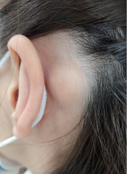
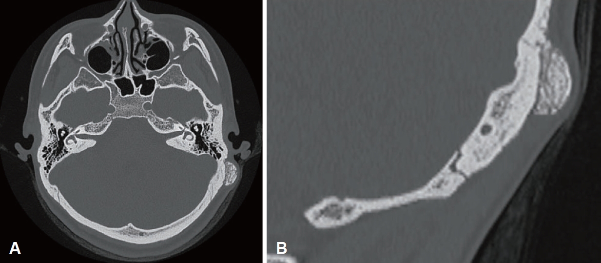
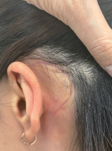
On physical examination, a firm solid mass measuring approximately 26×22 mm was palpable in the left retroauricular region (Fig. 1). On temporal bone CT, a round lesion measuring approximately 16×8 mm was observed in the left mastoid process of the temporal bone, and its density was similar to that of the adjacent cortical bone, suggesting a bony lesion rather than a soft tissue lesion. The lesion had a relatively well-defined margin and an isolated configuration that was distinguishable from the mastoid cortical bone, so based on these imaging findings, osteoma was suspected first (Fig. 2).
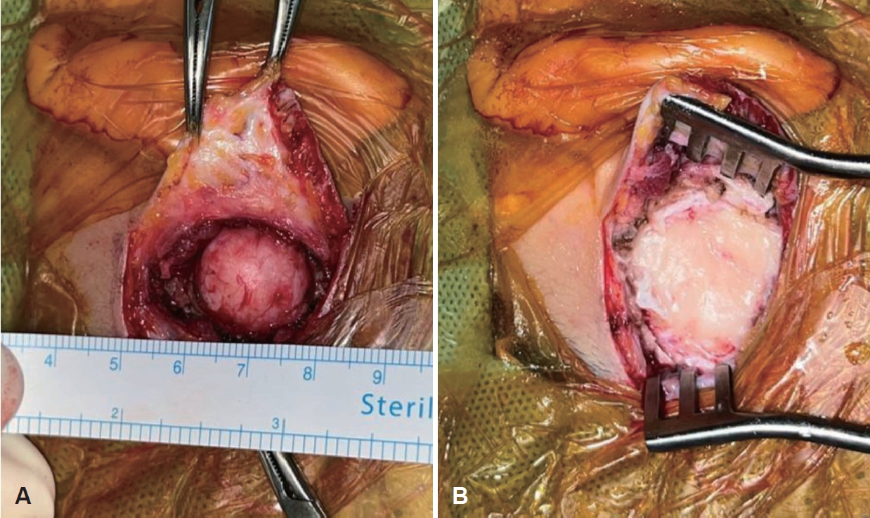
For definitive diagnosis, excisional biopsy was performed under local anesthesia. An incision was made and dissection was performed from the 12 o’clock to 7 o’clock direction centered on the lesion. Intraoperatively, the lesion was a firm bony mass that was dissected relatively well. The boundary between the mass and normal bone was relatively clear, and it was not firmly adherent to the temporal bone (Fig. 3A). Using a drill and gouge, the lesion was removed until normal cortical bone was clearly exposed (Fig. 3B). After confirming the absence of bleeding, closure and compressive dressing were applied, and the operation was completed.
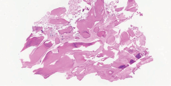
On histopathologic examination, irregularly arranged woven bone was observed within a fibrous stroma, and the final diagnosis was monostotic FD (Fig. 4). At 2 months after surgery, the surgical site recovered without specific complications (Fig. 5). At 1-year follow-up, there was no evidence of recurrence, and the patient is under outpatient observation.
Discussion
FD is a nonhereditary benign bone disease caused by abnormal differentiation of mesenchymal tissue, and it is often detected during adolescence as an asymptomatic or slowly progressive lesion [1,6]. Involvement of the temporal bone is reported to be uncommon even among craniofacial FD [2]. Lustig, et al. [7] reported temporal bone involvement in 5 of 21 patients (24%) with skull base FD. The proportions of monostotic and polyostotic types in temporal bone FD vary among studies. Some reports showed monostotic type in approximately 40% [2,7], whereas Frisch, et al. [8] reported approximately 70% as monostotic type. This difference is thought to result from differences in interpretation regarding whether a lesion that crosses cranial sutures to involve adjacent bones should be considered monostotic type or classified as polyostotic type.
Monostotic FD confined to the mastoid bone is extremely rare. Monini, et al. [5] reported one case of mastoid FD in a 39-year-old female who presented with headache. In Korea, there has been one report of FD involving the temporal bone accompanied by external auditory canal cholesteatoma [9], but monostotic FD confined to the mastoid antrum has not been reported.
Temporal bone FD may be accompanied by various symptoms depending on the location and extent of the lesion, including external auditory canal stenosis, conductive hearing loss, and cholesteatomatous otitis media [9]. According to previous reports, approximately 60% to 70% of patients with temporal bone FD have auditory symptoms [10], and Frisch, et al. [8] reported symptoms such as headache, hearing loss, and dizziness in 73% among 66 cases. However, as in this case, presentation only as a retroauricular mass without auditory symptoms is very rare, and differentiation from soft tissue masses such as lymphadenopathy, epidermoid cyst, and lipoma is required [11,12]. Bony lesions to be included in the differential diagnosis are osteoma, osteoblastoma, and FD [13].
Radiologically, FD is characterized by a ground-glass appearance on CT [14], but it may also appear as a homogeneous hyperdense lesion or a cystic lesion, which can make differential diagnosis difficult. Therefore, when imaging findings are unclear, confirmation through histopathologic examination is required. Histopathologically, irregularly arranged woven bone within a fibrous stroma is characteristic, and osteoblasts or osteoclasts are generally not observed [6]. Lesions to be differentiated include osteoma, osteoblastoma, and osteosarcoma. Osteoma is composed of mature lamellar bone. Osteoblastoma shows woven bone formation within a fibrovascular stroma and a distinct osteoblastic rimming layer at the periphery [13,14].
In this case, irregular woven bone was observed in a fibrous stroma. There was no osteoblastic rimming layer or invasive findings suggestive of osteosarcoma. The findings were also different from the mature lamellar bone features of osteoma, so FD could be confirmed.
Most cases of FD can be managed with observation. However, surgical excision is considered when there are cosmetic problems, functional abnormalities, or a need for differential diagnosis [8]. In this case, osteoma or a benign soft tissue lesion was suspected based on imaging and clinical findings, and excision allowed definitive diagnosis and treatment at the same time.
According to previous reports, in craniofacial FD, recurrence after complete excision is reported to be less than 10%, whereas recurrence after incomplete excision is reported to be approximately 40%, so long-term follow-up is necessary [15]. In this case, there has been no evidence of recurrence up to 1 year after complete excision, but periodic imaging and outpatient follow-up are planned to continue long-term observation for more than 5 years.
In conclusion, FD confined to the mastoid bone is rare, but when it presents as a retroauricular mass, it may be mistaken for a common soft tissue lesion. This case suggests that FD should be included in the differential diagnosis in such situations and may serve as a reference for clinical diagnosis and treatment decisions.