Introduction
Thyroid nodules are found in up to 50% of the adult population on routine ultrasound (US) examination [1,2]. For the evaluation of thyroid nodules, high-resolution US, with or without US-guided fine-needle aspiration cytology (FNAC), has been commonly used and is acknowledged as the first-line imaging modality [2,3]. Indeed, US is typically the only image used in evaluation of thyroid nodules; nearly all international guidelines for evaluation of thyroid nodules do not recommend use of other imaging studies, such as CT or MRI [2,4]. However, US examination is highly operator-dependent and provides narrow-view images that are restricted to the region where the probe contacts; these cannot show anatomical relationships of the entire neck within the same view. In addition, because the thyroid gland neighbors and abuts many structures (e.g., strap muscle, trachea, esophagus, parathyroid gland, and recurrent laryngeal nerve), perithyroidal pathologies arising from those structures may mimic thyroid pathologies when viewed with US alone.
Bronchogenic cysts are small, solitary cysts or sinuses that are formed from an abnormal budding of the tracheal diverticulum in the 16th week of gestation [5,6]. They are typically located in the mediastinum of pediatric patients, but may occasionally be found around the lower pole of the thyroid gland or level VI region, even in adults, during thyroid US [5-7]. However, US findings of cervical bronchogenic cyst have not yet been reported; thus, preoperative diagnosis is quite challenging, and may lead to unnecessary diagnostic surgery.
In these case reports, we describe US findings of bronchogenic cyst that may mimic papillary thyroid carcinoma and introduce some sources of diagnostic error, with the aim of improved understanding of cervical bronchogenic cyst, as well as avoidance of misdiagnosis as suspicious thyroid nodule.
Case
The US systems used in these cases study were the HS 70A (Samsung Medison, Seoul, Korea) and the HD7 (Philips Healthcare, Bothell, WA, USA), each with a high-frequency linear transducer (3-12 MHz). Both patients underwent contrast-enhanced CT with a slice thickness of 3.0 mm. The US findings were compared with CT findings and clinical records were reviewed for each patient.
Case 1
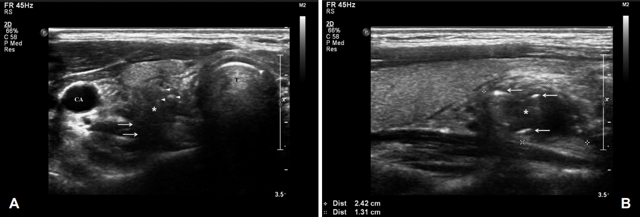
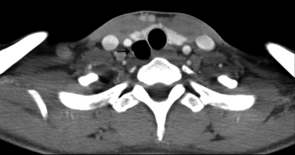
A 29-year-old female visited our clinic for further evaluation of a thyroid nodule detected on thyroid US during a wellness examination. She complained of a mild foreign body sensation in throat, but the symptom was not considered associated with the nodule. On US, an ill-defined hypoechoic nodule was identified at the inferior pole of the right thyroid gland. The nodule had several internal echogenic foci resembling microcalcification; posterior extrathyroidal extension was suspected (Fig. 1A). On longitudinal US view, several arc-shaped hyperechoic lines were present at the peripheral portion of the nodule (Fig. 1B). According to the sonographic patterns in the American Thyroid Association (ATA) guidelines, as well as several thyroid imaging reporting and data systems (TIRADS), the nodule was classified as highly suspicious; thus, FNAC was performed [2,8]. The nodule was categorized as benign; however, it showed numerous ciliated columnar epithelial cells rather than follicular cells. With these findings, we speculated that the lesion may have arisen from the respiratory tract; thus, we performed CT to characterize the anatomical relationship between the lesion and aerodigestive tract. The CT scan demonstrated an air-filled cyst at the posterior aspect of the right thyroid gland, which involved lower cervical to mediastinal regions (Fig. 2). Based on the results of US, FNAC, and CT analyses, the lesion was confirmed as a bronchogenic cyst. Because the patient had no symptoms related to the bronchogenic cyst, we recommended close observation with regular follow-up. For 35 months of follow-up, the lesion has remained stationary and the patient has not exhibited any complications.
Case 2
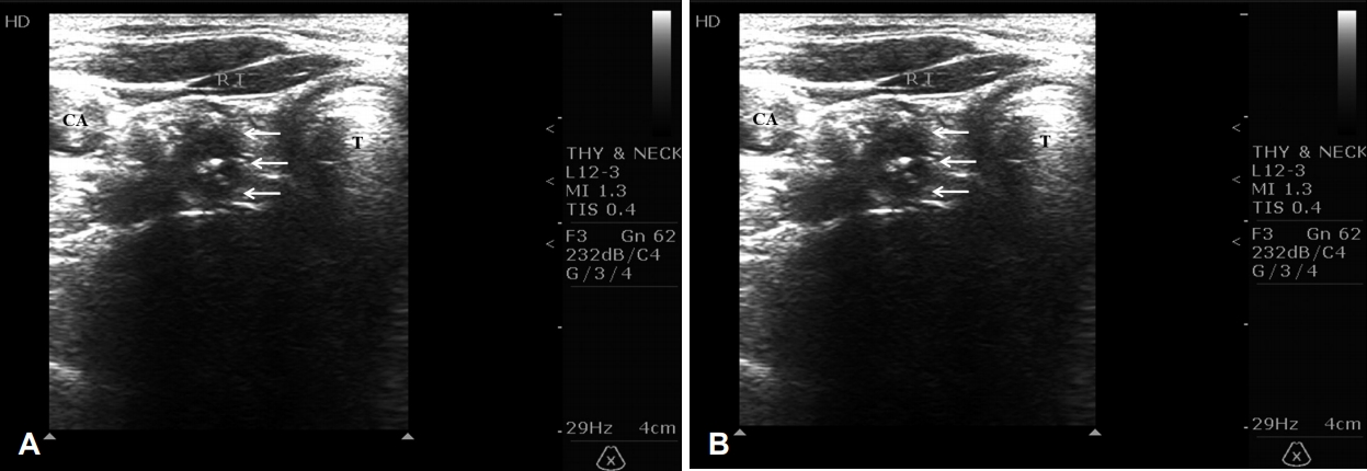
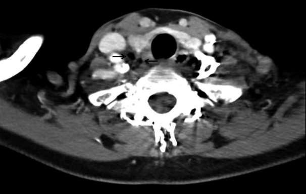
A 52-year-old female was transferred to our clinic for further evaluation of a thyroid nodule detected during a previous thyroid US. At the time of presentation, her chief complaint was mild odynophagia; laryngoscopic examination was unremarkable, with the exception of findings that indicated laryngopharyngeal reflux disease. On US, an ill-defined hypoechoic nodule was found at the inferior pole of the right thyroid gland (Fig. 3A) and right level VI region (Fig. 3B); it exhibited several internal echogenic foci mimicking calcification. According to the sonographic patterns of the ATA guidelines, as well as several TIRADS, thyroid cancer with central metastasis was included in the differential diagnosis [2,8]. However, considering our previous experience with a bronchogenic cyst, we recommended a CT scan prior to FNAC. On CT scan, several air-filled cysts were found, extending from the inferior pole of the right thyroid gland to the upper mediastinum (Fig. 4). There was no evidence of a suspicious thyroid nodule with calcification or central metastasis. Based on these findings, the lesion was diagnosed as a bronchogenic cyst. Because the patient had no symptoms related to the bronchogenic cyst, we recommended close observation with regular follow-up. For 30 months of followup, the lesion has remained stationary and the patient has not exhibited any complications.
Discussion
A bronchogenic cyst is a rare congenital anomaly formed by abnormal budding of the tracheal diverticulum; it is typically located in the mediastinum, most often in pediatric patients [5,6]. When it is located in the cervical region in adults, it is rarely diagnosed on preoperative US, due to its rarity and poor understanding of its US findings. Although there have been several reports of cervical bronchogenic cysts in adults, these were suspected to be thyroid tumor, parathyroid tumor, or brachial anomaly during initial preoperative evaluations; none were diagnosed without surgical intervention [5-7,9-11]. Furthermore, in most previous reports, US findings of bronchogenic cyst were not addressed.
In the two cases we have described, bronchogenic cysts appeared as ill-defined inhomogeneous hypoechoic nodules on US, exhibiting several echogenic foci or arc-shaped echogenic lines. Although these were designated as “cysts,” their capsules were not distinct. The clinical significance of such cysts on thyroid US is that they can mimic thyroid cancer of the lower pole of the thyroid gland and/or its central metastases, due to their hypoechogenicity, unclear or irregular margin, and echogenic foci, which may be misidentified as microcalcification; these findings correspond to the typical findings of a highly suspicious thyroid nodule [2,8].
Several clues can be used to distinguish bronchogenic cysts from true thyroid malignancies. First, on US, echogenic foci or arc-shaped lines of the lesion do not exhibit clean acoustic shadowing, (i.e., a uniformly anechoic signal behind a structure) indicative of calcification; rather, they may demonstrate posterior reverberation and dirty acoustic shadowing (i.e., low-level echoes in the shadow, deep to air) [12,13]. In addition, when needle aspiration is performed, the tactile sense of the needle is quite different from that of thyroid malignancy, because the internal contents of bronchogenic cysts constitute air without solid components; in contrast, these solid components are present in thyroid cancer. With respect to cytology, because bronchogenic cysts are lined by respiratory-type epithelium, which is characterized by cilia, FNAC can demonstrate ciliated columnar epithelial cells; these suggest that the lesion developed from the respiratory tract. However, obtaining an adequate specimen is often difficult, because this is an air-filled cyst composed of various fluids; therefore, preoperative diagnosis is difficult. Further, inappropriate FNAC can cause cyst rupture, pneumomediastinum, and secondary infection. Therefore, if a bronchogenic cyst is suspected on US, an additional CT scan can be helpful for diagnosis in that it demonstrates an air-filled cyst involving the lower cervical region around the thyroid gland; this may allow avoidance of unnecessary invasive procedures. However, confirmation of the diagnosis should be based on pathological examination demonstrating a cystic lesion lined with respiratory-type epithelium (ciliated columnar epithelial cells) [10,11].
In addition to thyroid cancer, US findings of bronchogenic cyst can mimic those of esophageal diverticulum, which exhibits similar anatomical location and the presence of air, as in bronchogenic cysts. However, US findings of diverticulum involve changes in the shape and shadowing from microbubbles entering the diverticulum during swallowing, connection to the esophagus, a peripheral echogenic line, and a boundary hypoechoic zone. These findings facilitate differentiation of esophageal diverticulum from other disease entities [14,15].
Regarding air-filled structures, such as bronchogenic cysts, the US could not clearly depict the margin of the lesion; thus, image discordance might be observed between the US and CT scans. This is an important limitation of the use of US images to examine air-filled structures. This phenomenon is regarded as a type of “dirty shadowing” artifact, which occurs as a results of the high degree of reflection at air-tissue interfaces [12,13]. Because the energy of a sound pulse reflected off air is similar that of the transmitted pulse, the reflected pulse will interact with interfaces in front of the air; this interaction produces secondary reflections, which travel back to the air surface and are reflected this surface back to the transducer. Notably, these secondary reflections produce low-level echoes in the shadow deep to the air, thereby causing the “dirty” appearance. Consequently, structures posterior to the large air-filled structure cannot be depicted clearly by US, and may observed as distorted images that are not representative of the true anatomy [12,13]. US operator should recognize this phenomenon caused by dirty shadowing artifacts from air, in order to avoid misinterpretation of US images when examining air-filled lesions.
In conclusion, our cases showed that cervical bronchogenic cysts can present as highly suspicious thyroid nodules on thyroid US. However, recognition of this disease entity and comprehensive understanding of its presentation on US can facilitate appropriate diagnosis and avoid unnecessary diagnostic surgery.